 Back
BackFunctional Anatomy of Prokaryotic and Eukaryotic Cells: Structure and Function
Study Guide - Smart Notes
 Tailored notes based on your materials, expanded with key definitions, examples, and context.
Functional Anatomy of Prokaryotic and Eukaryotic Cells
Overview: Prokaryotic vs. Eukaryotic Cells
This section compares the fundamental differences between prokaryotic and eukaryotic cells, which are the two primary cell types in microbiology. Understanding these differences is essential for classifying microorganisms and predicting their cellular functions.
Prokaryotes: Characterized by a single, circular chromosome not enclosed in a membrane, absence of histones and organelles, peptidoglycan cell walls (in bacteria), and division by binary fission.
Eukaryotes: Possess paired chromosomes within a nuclear membrane, contain histones and organelles, may have polysaccharide cell walls, and divide by mitosis.
Shape and Arrangement of Bacterial Cells
Common Bacterial Shapes
Bacteria exhibit a variety of shapes, which are important for identification and classification. The main shapes include:
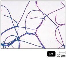
Bacillus: Rod-shaped
Coccus: Spherical-shaped
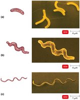
Spiral Forms: Includes vibrio (curved rods), spirillum (rigid spirals), and spirochete (flexible spirals)
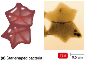
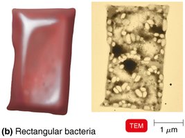
Unusual Shapes: Star-shaped and rectangular bacteria are less common but notable for their unique morphology.
Arrangements of Bacterial Cells
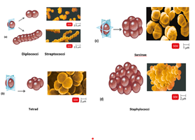
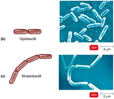
Bacterial cells can be arranged in characteristic patterns due to their division planes:
Pairs: Diplococci, diplobacilli
Chains: Streptococci, streptobacilli
Clusters: Staphylococci
Groups of Four: Tetrads
Cubelike Groups of Eight: Sarcinae
Structure of a Prokaryotic Cell
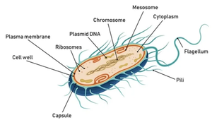
Generalized Structure
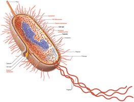
Prokaryotic cells have a simple structure but contain all the essential components for life. Key features include the cell wall, plasma membrane, cytoplasm, nucleoid, ribosomes, and various surface structures.
Surface Structures of Prokaryotic Cells
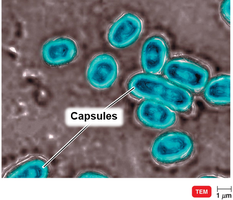
Glycocalyx
The glycocalyx is a viscous, gelatinous layer external to the cell wall, composed of polysaccharide and/or polypeptide. It exists in two forms:
Capsule: Neatly organized and firmly attached to the cell wall.
Slime Layer: Unorganized and loosely attached.
Functions include protection from phagocytosis, contribution to virulence, and facilitation of biofilm formation.
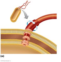
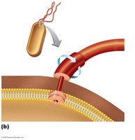
Flagella
Flagella are long, filamentous appendages used for motility. They are composed of the protein flagellin and have three main parts:
Filament: Outermost region
Hook: Connects filament to basal body
Basal Body: Anchors flagellum to cell wall and membrane; structure differs between Gram-positive and Gram-negative bacteria
Flagella arrangements include monotrichous (single), lophotrichous (tufts), amphitrichous (both ends), and peritrichous (all over surface).
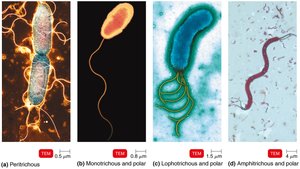
Flagella enable movement toward or away from stimuli (taxis) and are important antigens (H antigens).
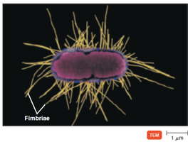
Fimbriae and Pili
Fimbriae are short, hairlike appendages that allow for attachment to surfaces. Pili are longer, fewer in number, and involved in motility (gliding, twitching) and DNA transfer (conjugation).
The Bacterial Cell Wall
Functions and Composition
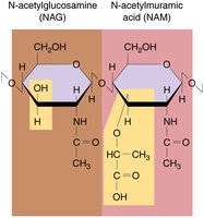
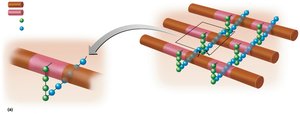
The bacterial cell wall provides structural support, prevents osmotic lysis, and contributes to pathogenicity. It is primarily composed of peptidoglycan, a polymer of repeating disaccharides (N-acetylglucosamine [NAG] and N-acetylmuramic acid [NAM]) linked by polypeptides.
Gram-Positive Cell Walls
Gram-positive bacteria have thick peptidoglycan layers and teichoic acids, which link the cell wall to the plasma membrane and provide antigenic specificity. The structure includes peptide cross-bridges and tetrapeptide side chains.
Teichoic acids carry a negative charge and regulate movement of cations.
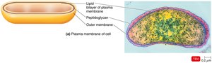
Gram-Negative Cell Walls
Gram-negative bacteria have a thin peptidoglycan layer and an outer membrane composed of lipopolysaccharides (LPS), lipoproteins, and phospholipids. The LPS contains:
O polysaccharide: Functions as an antigen
Lipid A: Acts as an endotoxin
Porins: Proteins that form channels through the membrane
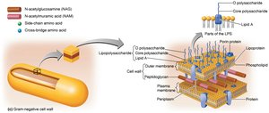
The outer membrane protects against phagocytes, complement, and antibiotics.
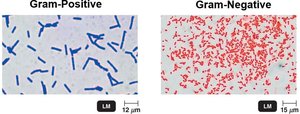
Gram Stain Mechanism
The Gram stain differentiates bacteria based on cell wall structure:
Gram-positive: Alcohol dehydrates peptidoglycan, trapping crystal violet-iodine complexes (cells appear purple).
Gram-negative: Alcohol dissolves outer membrane and leaves holes in peptidoglycan, allowing dye to wash out (cells appear colorless until counterstained with safranin, then appear pink/red).
Atypical Cell Walls
Some bacteria, such as Mycobacterium and Nocardia, have acid-fast cell walls containing mycolic acid, a waxy lipid bound to peptidoglycan. These stain with carbolfuchsin.
The Plasma (Cytoplasmic) Membrane
Structure and Function
The plasma membrane is a phospholipid bilayer with embedded proteins, enclosing the cytoplasm. It is selectively permeable, allowing passage of certain molecules while restricting others. The membrane contains enzymes for ATP production and, in some bacteria, photosynthetic pigments on infoldings called chromatophores.
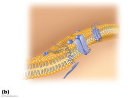
Damage to the membrane by alcohols, detergents, or antibiotics can cause leakage of cell contents and cell death.
Summary Table: Comparison of Gram-Positive and Gram-Negative Bacteria
Feature | Gram-Positive | Gram-Negative |
|---|---|---|
Peptidoglycan Layer | Thick | Thin |
Teichoic Acids | Present | Absent |
Outer Membrane | Absent | Present |
Lipopolysaccharide (LPS) | Absent | Present |
Sensitivity to Penicillin | High | Low |
Gram Stain Color | Purple | Pink/Red |
