 Back
Functional Anatomy of Prokaryotic and Eukaryotic Cells: Structure and Function
Study Guide - Smart Notes
 Tailored notes based on your materials, expanded with key definitions, examples, and context.
Comparing Prokaryotic and Eukaryotic Cells
Overview
Prokaryotic and eukaryotic cells are the two fundamental types of cells that make up all living organisms. Understanding their similarities and differences is essential for microbiology, as it provides the foundation for studying microbial structure, function, and classification.
Prokaryotic cells lack a membrane-bound nucleus and organelles. Their genetic material is found in a single, circular chromosome.
Eukaryotic cells possess a true nucleus enclosed by a nuclear membrane and contain various membrane-bound organelles.
Both cell types share basic chemical composition and metabolic pathways but differ in complexity and organization.
Key Differences:
Feature | Prokaryotes | Eukaryotes |
|---|---|---|
Nucleus | Absent | Present |
Chromosomes | Single, circular | Multiple, linear |
Cell Wall | Peptidoglycan (Bacteria), Pseudomurein (Archaea) | Cellulose (plants), Chitin (fungi), or absent |
Organelles | Absent | Present |
Division | Binary fission | Mitosis/meiosis |

The Prokaryotic Cell
Size, Shape, and Arrangement
Bacteria exhibit a variety of shapes and arrangements, which are important for identification and classification.
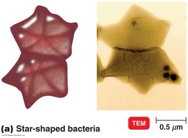
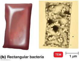
Shapes: Coccus (spherical), Bacillus (rod-shaped), Spiral (vibrio, spirillum, spirochete), Star-shaped, Rectangular
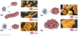
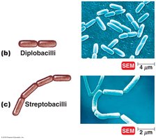
Arrangements: Diplococci (pairs), Streptococci (chains), Staphylococci (clusters), Tetrads (groups of four), Sarcinae (cubical groups of eight)
Pleomorphism: Some bacteria can vary in shape.
Structures External to the Cell Wall
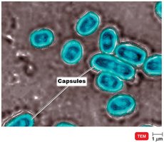
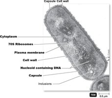
Glycocalyx: A gelatinous polysaccharide/polypeptide layer external to the cell wall. It can be a capsule (organized, firmly attached) or a slime layer (unorganized, loose).
Functions: Protection from phagocytosis, adherence to surfaces, prevention of desiccation, and contribution to virulence.
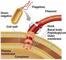
Flagella: Long, whip-like appendages used for motility. Composed of filament, hook, and basal body. Movement is by rotation.
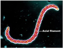
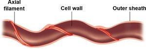
Axial filaments: Found in spirochetes, these are internal flagella that enable corkscrew movement.
Fimbriae: Short, hairlike structures for attachment.
Pili: Longer than fimbriae; involved in motility and DNA transfer (conjugation).
The Cell Wall
The bacterial cell wall is a rigid structure that provides shape, protection, and prevents osmotic lysis. It is a key target for antibiotics.
Peptidoglycan: Main component in bacteria, consisting of repeating units of N-acetylglucosamine (NAG) and N-acetylmuramic acid (NAM) linked by polypeptides.
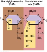
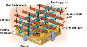
Gram-positive cell walls: Thick peptidoglycan, teichoic acids, high susceptibility to penicillin, disrupted by lysozyme.
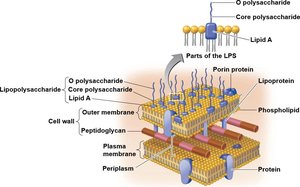
Gram-negative cell walls: Thin peptidoglycan, outer membrane with lipopolysaccharide (LPS), periplasmic space, low susceptibility to penicillin.
Special Cases:
Mycoplasma: Lack cell walls.
Archaea: Have pseudomurein, not peptidoglycan.
Acid-fast bacteria: Have mycolic acid outside a thin peptidoglycan layer.
Structures Internal to the Cell Wall
Plasma (Cytoplasmic) Membrane: Phospholipid bilayer with proteins, selectively permeable, site of metabolic enzymes.
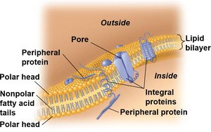
Cytoplasm: Mostly water, contains proteins, carbohydrates, lipids, ions, DNA, ribosomes, inclusions, and cytoskeleton proteins.
Nucleoid: Region containing the bacterial chromosome (DNA) and sometimes plasmids.
Ribosomes: Sites of protein synthesis, 70S type (50S + 30S subunits).
Inclusions: Reserve deposits (e.g., metachromatic granules, glycogen, lipid inclusions, sulfur granules, magnetosomes, gas vacuoles).
Endospores: Resting structures for survival in harsh conditions.
The Eukaryotic Cell
General Structure
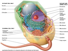
Eukaryotic cells are more complex than prokaryotic cells and contain a variety of membrane-bound organelles.
Organisms: Protozoa, algae, fungi, plants, animals
Key features: Nucleus, endoplasmic reticulum, Golgi apparatus, mitochondria, chloroplasts (in plants/algae), lysosomes, vacuoles, cytoskeleton
Cell Wall and Glycocalyx
Cell wall: Present in plants (cellulose), fungi (chitin), and some protists; absent in animals.
Glycocalyx: Carbohydrate-rich layer found in animal cells, involved in cell recognition and attachment.
Plasma (Cytoplasmic) Membrane
Phospholipid bilayer with proteins, sterols, and carbohydrates.
Functions: Selective permeability, transport, cell signaling, endocytosis (phagocytosis, pinocytosis).
Ribosomes
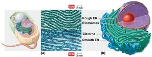
80S ribosomes: Found in cytoplasm and on rough ER (60S + 40S subunits).
70S ribosomes: Found in mitochondria and chloroplasts, similar to prokaryotic ribosomes.
Organelles
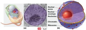
Nucleus: Double membrane, contains DNA as chromatin/chromosomes, site of transcription.
Endoplasmic Reticulum (ER): Rough ER (with ribosomes, protein synthesis), Smooth ER (lipid synthesis, detoxification).
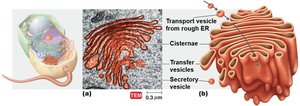
Golgi Complex: Modifies, sorts, and packages proteins and lipids for secretion or delivery to other organelles.
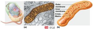
Mitochondria: Sites of ATP production via cellular respiration; contain their own DNA and 70S ribosomes.
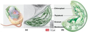
Chloroplasts: Sites of photosynthesis in plants and algae; contain thylakoids, DNA, and 70S ribosomes.
Lysosomes: Contain digestive enzymes for breakdown of macromolecules.
Vacuoles: Storage and structural support, especially in plant cells.
Endosymbiotic Theory
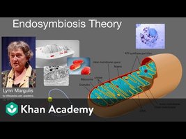
The endosymbiotic theory proposes that eukaryotic organelles such as mitochondria and chloroplasts originated from free-living prokaryotes that were engulfed by ancestral eukaryotic cells. Evidence includes the presence of 70S ribosomes and circular DNA in these organelles, and their division by binary fission.
Summary Table: Prokaryotic vs. Eukaryotic Cells
Feature | Prokaryotic Cell | Eukaryotic Cell |
|---|---|---|
Nucleus | No | Yes |
Organelles | No | Yes |
Cell Wall | Peptidoglycan (Bacteria), Pseudomurein (Archaea) | Cellulose (plants), Chitin (fungi), or absent |
Ribosomes | 70S | 80S (cytoplasm/ER), 70S (mitochondria/chloroplasts) |
Division | Binary fission | Mitosis/meiosis |
Key Terms and Concepts
Peptidoglycan: A polymer of sugars and amino acids forming the bacterial cell wall.
Endocytosis: Process by which eukaryotic cells engulf material from their environment.
Flagella: Structures for motility in both prokaryotes and eukaryotes, but with different structures and mechanisms.
Endosymbiotic theory: Hypothesis explaining the origin of mitochondria and chloroplasts in eukaryotic cells.
