 Back
BackFunctional Anatomy of Prokaryotic and Eukaryotic Cells: Structure, Function, and Comparison
Study Guide - Smart Notes
 Tailored notes based on your materials, expanded with key definitions, examples, and context.
Comparing Prokaryotic and Eukaryotic Cells
Overview of Cell Types
Prokaryotic and eukaryotic cells are the two fundamental cell types in microbiology. Their structural and functional differences are essential for understanding microbial physiology and classification.
Prokaryotes: Cells lacking a membrane-bound nucleus and organelles. Includes Bacteria and Archaea.
Eukaryotes: Cells with a true nucleus and membrane-bound organelles. Includes Fungi, Algae, Protozoa, and Helminths.
Key Differences:
Prokaryotes: Usually one circular chromosome, no histones, no organelles, peptidoglycan (bacteria) or pseudomurein (archaea) cell walls, divide by binary fission.
Eukaryotes: Paired chromosomes in nuclear membrane, histones, organelles, polysaccharide cell walls (when present), divide by mitosis.
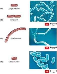
The Size, Shape, and Arrangement of Bacterial Cells
Bacterial Morphology
Bacteria exhibit a variety of shapes and arrangements, which are important for identification and classification.
Average size: 0.2–2.0 μm diameter, 2–8 μm length
Monomorphic: Most bacteria have a single, consistent shape.
Pleomorphic: Some bacteria can vary in shape.
Common Shapes:
Bacillus: Rod-shaped
Coccus: Spherical-shaped
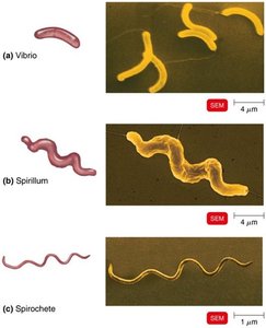
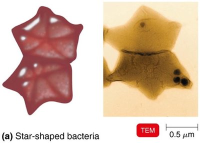
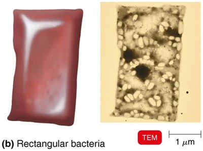
Spiral: Includes vibrio (curved rod), spirillum (rigid spiral), and spirochete (flexible spiral)
Star-shaped and rectangular forms also exist
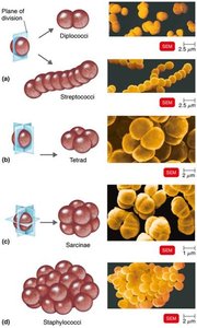
Arrangements:
Pairs: Diplococci, diplobacilli
Clusters: Staphylococci
Chains: Streptococci, streptobacilli
Groups of four: Tetrads
Cubelike groups of eight: Sarcinae
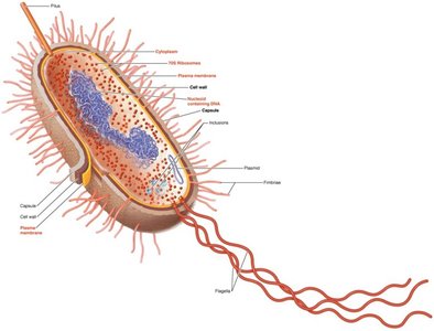
Structure of a Prokaryotic Cell
Cell Components
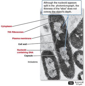
Prokaryotic cells contain several key structures, each with specific functions.
Capsule: Protective layer outside the cell wall
Cell wall: Provides structural support and shape
Plasma membrane: Controls entry and exit of substances
Cytoplasm: Site of metabolic activity
Nucleoid: Region containing DNA
Plasmid: Small, circular DNA molecules
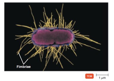
Fimbriae: Attachment structures
Pilus: Used for DNA transfer
Flagella: Motility structures
Inclusions: Storage granules
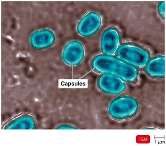
Glycocalyx
Structure and Function
The glycocalyx is an external, viscous, gelatinous layer made of polysaccharide and/or polypeptide. It exists in two forms:
Capsule: Neatly organized and firmly attached
Slime layer: Unorganized and loose
Functions:
Contributes to virulence by preventing phagocytosis and aiding adherence
Helps form biofilms, protecting cells and aiding attachment
Flagella, Axial Filaments, Fimbriae, and Pili
Flagella
Flagella are filamentous appendages that propel bacteria. They consist of three parts:
Filament: Outermost region
Hook: Attaches filament to basal body
Basal body: Anchors flagellum to cell wall and membrane
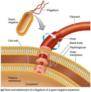
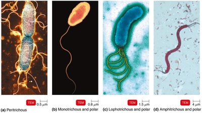
Flagella arrangements vary:
Peritrichous: Flagella all over the cell
Monotrichous: Single flagellum at one end
Lophotrichous: Multiple flagella at one end
Amphitrichous: Flagella at both ends
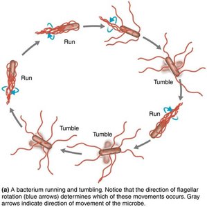
Flagella allow movement toward or away from stimuli (taxis) and rotate to produce "run" or "tumble" movements.
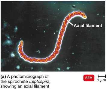
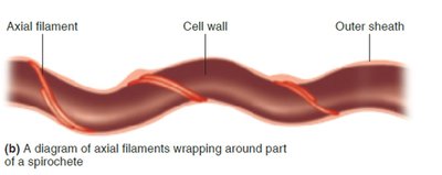
Axial Filaments
Axial filaments, also called endoflagella, are found in spirochetes. They are anchored at one end and cause the cell to move in a corkscrew manner.
Fimbriae and Pili
Fimbriae are hairlike appendages that allow for attachment and biofilm formation. Pili are involved in motility and DNA transfer (conjugation).
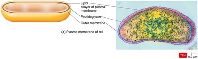
The Cell Wall
Structure and Function
The bacterial cell wall prevents osmotic lysis, protects the cell membrane, and contributes to pathogenicity. It is a site of action for antibiotics and is used to differentiate major groups of bacteria.
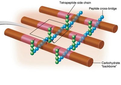
Peptidoglycan Structure
Peptidoglycan is a polymer of repeating disaccharides (N-acetylglucosamine and N-acetylmuramic acid) linked by polypeptides, forming a lattice structure.
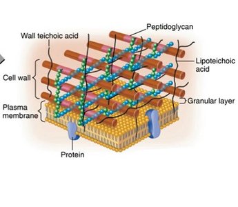
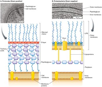
Gram-Positive Cell Walls
Gram-positive bacteria have thick peptidoglycan layers and teichoic acids, which regulate cation movement and provide antigenic specificity.
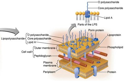
Gram-Negative Cell Walls
Gram-negative bacteria have thin peptidoglycan, a periplasmic space, and an outer membrane containing lipopolysaccharide (LPS), lipoproteins, and phospholipids. The outer membrane protects from phagocytes and antibiotics.
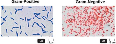
Gram Stain Mechanism
The Gram stain differentiates bacteria based on cell wall structure:
Gram-positive: Alcohol dehydrates peptidoglycan; crystal violet-iodine complexes remain.
Gram-negative: Alcohol dissolves outer membrane; complexes wash out; safranin stains cells.
Atypical Cell Walls
Some bacteria have atypical cell walls:
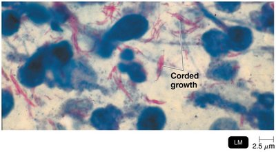
Acid-fast: Thick peptidoglycan with waxy mycolic acid; stains with carbolfuchsin.
Mycoplasmas: Lack cell walls; sterols in plasma membrane.
Archaea: Wall-less or walls of pseudomurein.
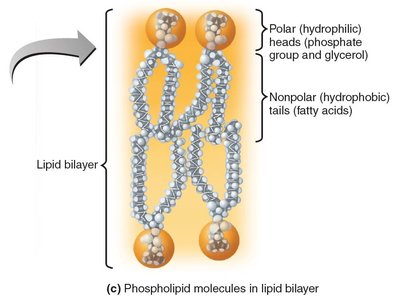
The Plasma (Cytoplasmic) Membrane
Structure
The plasma membrane is a phospholipid bilayer with peripheral, integral, and transmembrane proteins. Some proteins form channels; glycoproteins and glycolipids are present.
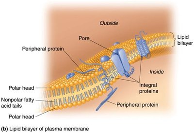
Function
The plasma membrane is selectively permeable, contains enzymes for ATP production, and may have photosynthetic pigments on foldings called chromatophores.
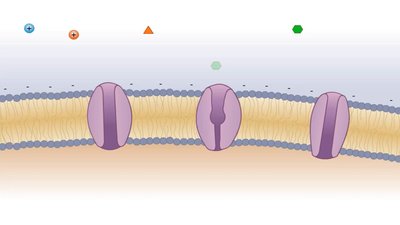
Movement of Materials Across Membranes
Passive Processes
Substances move from high to low concentration without energy expenditure.
Simple diffusion: Movement of solute to equilibrium.
Facilitated diffusion: Transport via membrane proteins.
Osmosis: Net movement of water across membrane.
Active Processes
Substances move from low to high concentration with energy expenditure.
Active transport: Requires transporter protein and ATP.
Group translocation: Substance is chemically altered during transport.
Cytoplasm, Nucleoid, Ribosomes, and Inclusions
Cytoplasm
The cytoplasm is a thick, aqueous, elastic substance inside the plasma membrane, containing DNA, ribosomes, and inclusions. The cytoskeleton aids in cell division, shape, growth, and DNA movement.
Nucleoid
The nucleoid contains the bacterial chromosome (circular, double-stranded DNA) and plasmids (extrachromosomal DNA).
Ribosomes
Sites of protein synthesis, composed of protein and ribosomal RNA. Prokaryotic ribosomes are 70S (50S + 30S subunits).
Inclusions
Reserve deposits in the cytoplasm, including:
Metachromatic granules (phosphate reserves)
Polysaccharide granules (energy reserves)
Lipid inclusions (energy reserves)
Sulfur granules (energy reserves)
Carboxysomes (CO2 fixation)
Gas vacuoles (buoyancy)
Magnetosomes (iron oxide inclusions)
Endospores
Formation and Function
Endospores are resting cells produced when nutrients are depleted. They are resistant to desiccation, heat, chemicals, and radiation, and can survive for thousands of years. Sporulation is endospore formation; germination is return to vegetative state.
Comparing Eukaryotic and Prokaryotic Structures
Flagella and Cilia
Eukaryotic flagella and cilia are projections used for locomotion or moving substances. Flagella are long and few; cilia are short and numerous. Both consist of microtubules in a 9+2 array and move in a wavelike manner.
Cell Wall and Glycocalyx
Eukaryotic cell walls are found in plants, algae, and fungi, made of carbohydrates (cellulose, chitin, glucan, mannan). Glycocalyx is found in animal cells, strengthens the cell surface, aids attachment, and is involved in cell–cell recognition.
Plasma Membrane
Eukaryotic plasma membranes are similar to prokaryotic ones but contain sterols and carbohydrates for attachment and recognition. Functions include selective permeability, diffusion, osmosis, active transport, and endocytosis (phagocytosis, pinocytosis, receptor-mediated).
Cytoplasm
Eukaryotic cytoplasm contains cytosol, cytoskeleton (microfilaments, intermediate filaments, microtubules), and exhibits cytoplasmic streaming.
Ribosomes
Eukaryotic ribosomes are 80S (60S + 40S subunits), found free in cytoplasm or bound to endoplasmic reticulum. 70S ribosomes are found in mitochondria and chloroplasts.
The Evolution of Eukaryotes
Endosymbiotic Theory
The endosymbiotic theory explains the origin of eukaryotes: larger bacterial cells engulfed smaller ones, forming organelles like mitochondria and chloroplasts. Evidence includes similarities in size, shape, DNA, reproduction, ribosomes, and double membranes between these organelles and bacteria.
----------------------------------------
