 Back
BackFunctional Anatomy of Prokaryotic and Eukaryotic Cells: Structure, Function, and Comparison
Study Guide - Smart Notes
 Tailored notes based on your materials, expanded with key definitions, examples, and context.
Comparing Prokaryotic and Eukaryotic Cells
Overview of Cell Types
Prokaryotic and eukaryotic cells represent two fundamental organizational types in microbiology. Understanding their structural and functional differences is essential for grasping microbial physiology and taxonomy.
Prokaryotes: Cells lacking a membrane-bound nucleus; include Bacteria and Archaea.
Eukaryotes: Cells with a true nucleus and membrane-bound organelles; include plants, animals, fungi, and protists.
Key Differences: Chromosome structure, presence of histones, organelles, cell wall composition, and division mechanisms.
Example: Bacteria (prokaryotes) divide by binary fission, while eukaryotic cells divide by mitosis.
The Size, Shape, and Arrangement of Bacterial Cells
Bacterial Morphology
Bacteria exhibit a variety of shapes and arrangements, which are important for identification and classification.
Average Size: 0.2–2.0 μm diameter, 2–8 μm length.
Monomorphic: Most bacteria have a single, consistent shape.
Pleomorphic: Some bacteria can vary in shape.
Main Shapes
Bacillus: Rod-shaped
Coccus: Spherical-shaped
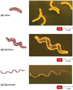
Spiral: Includes Vibrio (curved rod), Spirillum (rigid spiral), Spirochete (flexible spiral)
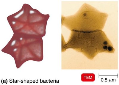
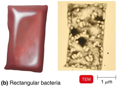
Star-shaped and Rectangular forms (rare)
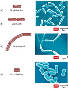
Arrangements of Cocci and Bacilli
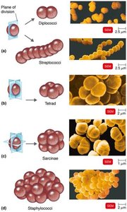
Bacterial cells can be arranged in characteristic patterns based on their division planes.
Cocci: Diplococci (pairs), Streptococci (chains), Tetrads (groups of four), Sarcinae (cubelike groups of eight), Staphylococci (clusters)
Bacilli: Single, Diplobacilli (pairs), Streptobacilli (chains), Coccobacilli (short rods)
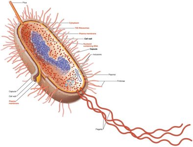
Structure of a Prokaryotic Cell
Cell Components
Prokaryotic cells contain essential structures for survival and reproduction. Not all bacteria possess every structure.
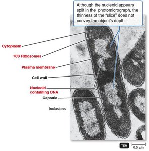
Capsule: Protective outer layer
Cell wall: Provides shape and protection
Plasma membrane: Controls entry and exit of substances
Cytoplasm: Site of metabolic activity
70S ribosomes: Protein synthesis
Nucleoid: Contains DNA
Plasmid: Extra-chromosomal DNA
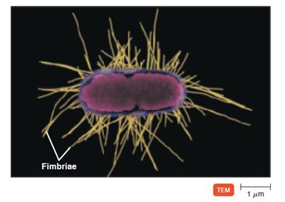
Fimbriae: Attachment structures
Pilus: DNA transfer and motility
Flagella: Motility
Inclusions: Storage granules
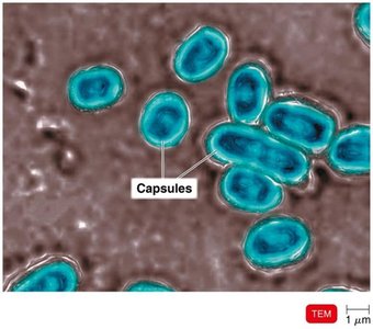
Glycocalyx
Structure and Function
The glycocalyx is an external, viscous, gelatinous layer composed of polysaccharides and/or polypeptides. It exists in two forms:
Capsule: Neatly organized, firmly attached
Slime layer: Unorganized, loose
Functions:
Contributes to virulence by preventing phagocytosis and aiding adherence
Helps form biofilms, protecting cells and aiding attachment
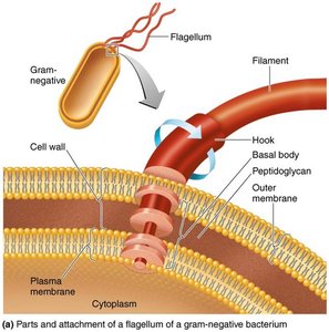
Flagella, Axial Filaments, Fimbriae, and Pili
Flagella
Flagella are filamentous appendages that propel bacteria. They consist of three parts: filament, hook, and basal body. Bacterial flagella are powered by a proton gradient.
Filament: Outermost region
Hook: Attaches filament to basal body
Basal body: Anchors flagellum to cell wall and membrane
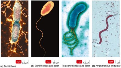
Flagellar Arrangements
Bacteria exhibit various flagellar arrangements, which aid in motility and identification.
Peritrichous: Flagella all over the cell
Monotrichous: Single flagellum at one end
Lophotrichous: Multiple flagella at one end
Amphitrichous: Flagella at both ends
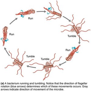
Flagellar Movement
Flagella rotate to produce "runs" (straight movement) and "tumbles" (change in direction). Flagella proteins serve as H antigens for serovar identification.
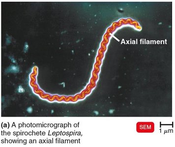
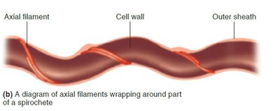
Axial Filaments (Endoflagella)
Axial filaments are found in spirochetes and are anchored at one end of the cell. Their rotation causes the cell to move in a corkscrew fashion.
Fimbriae and Pili
Fimbriae are hairlike appendages that allow for attachment and biofilm formation. Pili are involved in motility and DNA transfer (conjugation).
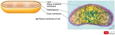
The Cell Wall
Structure and Function
The bacterial cell wall prevents osmotic lysis, protects the cell membrane, and contributes to pathogenicity. It is a site of action for antibiotics and is composed of peptidoglycan.
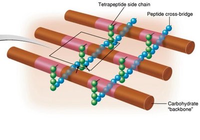
Peptidoglycan: Polymer of repeating disaccharides (N-acetylglucosamine and N-acetylmuramic acid) linked by polypeptides, forming a lattice structure.
Penicillin: Interferes with peptide cross-bridges, weakening the cell wall.
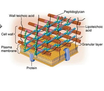
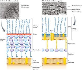
Gram-Positive Cell Walls
Gram-positive bacteria have thick peptidoglycan layers and teichoic acids, which regulate cation movement and provide antigenic specificity.
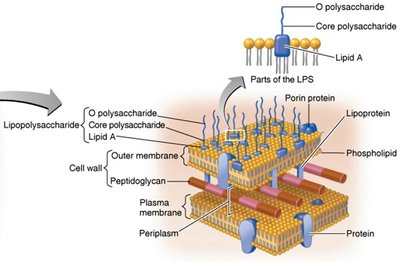
Gram-Negative Cell Walls
Gram-negative bacteria have thin peptidoglycan, an outer membrane with lipopolysaccharide (LPS), lipoproteins, and phospholipids. The outer membrane protects from phagocytes and antibiotics.
LPS: O polysaccharide (antigen), Lipid A (endotoxin)
Porins: Channels for molecule passage
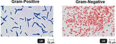
Gram Stain Mechanism
The Gram stain differentiates bacteria based on cell wall structure:
Gram-positive: Alcohol dehydrates peptidoglycan; crystal violet-iodine complex remains.
Gram-negative: Alcohol dissolves outer membrane; complex washes out; safranin stains cells.
Atypical Cell Walls
Some bacteria have atypical cell walls:
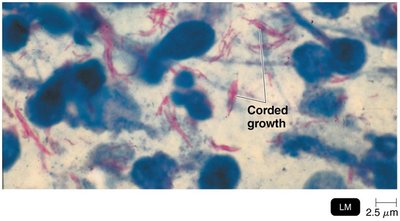
Acid-fast: Thick peptidoglycan, waxy mycolic acid; stain with carbolfuchsin.
Mycoplasmas: Lack cell walls; sterols in membrane.
Archaea: Wall-less or walls of pseudomurein (lack NAM and D-amino acids).
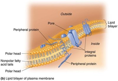
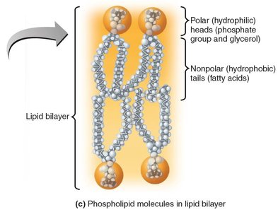
The Plasma (Cytoplasmic) Membrane
Structure
The plasma membrane is a phospholipid bilayer enclosing the cytoplasm, with peripheral, integral, and transmembrane proteins. Some proteins form channels; glycoproteins and glycolipids are present.
Function
Selective permeability: Allows passage of some molecules
ATP production: Contains enzymes for energy generation
Photosynthetic pigments: On foldings called chromatophores
Movement of Materials Across Membranes
Passive Processes
Simple diffusion: Movement from high to low concentration
Facilitated diffusion: Uses transporter proteins for ions and larger molecules
Osmosis: Net movement of water across a selectively permeable membrane
Osmotic pressure: Pressure needed to stop water movement
Active Processes
Active transport: Requires transporter protein and ATP; moves substances against gradient
Group translocation: Requires transporter protein and PEP; substance is chemically altered during transport
Cytoplasm, Nucleoid, Ribosomes, and Inclusions
Cytoplasm
The cytoplasm is a thick, aqueous, elastic substance inside the plasma membrane, containing DNA, ribosomes, and inclusions. The cytoskeleton aids in cell division, shape, growth, and DNA movement.
Nucleoid
Bacterial chromosome: Circular, double-stranded DNA
Plasmids: Small, extrachromosomal DNA circles; carry non-crucial genes
Ribosomes
Sites of protein synthesis
70S: 50S large + 30S small subunits
Antibiotics: Streptomycin, gentamicin, erythromycin, chloramphenicol target prokaryotic ribosomes
Inclusions
Metachromatic granules: Phosphate reserves
Polysaccharide granules: Energy reserves
Lipid inclusions: Energy reserves
Sulfur granules: Energy reserves
Carboxysomes: RuBisCO enzyme for CO2 fixation
Gas vacuoles: Buoyancy
Magnetosomes: Iron oxide inclusions
Endospores
Formation and Function
Endospores are resting cells produced by certain bacteria when nutrients are depleted. They are highly resistant to desiccation, heat, chemicals, and radiation, and can survive for thousands of years. Endospore formation (sporulation) is a survival mechanism, not a reproductive process.
Produced by: Bacillus and Clostridium
Germination: Endospore returns to vegetative state
Comparing Eukaryotic and Prokaryotic Structures
Flagella and Cilia
Eukaryotic flagella and cilia are projections used for locomotion or moving substances. Flagella are long and few; cilia are short and numerous. Both consist of microtubules arranged in a 9+2 array and move in a wavelike manner.
Cell Wall and Glycocalyx
Cell wall: Found in plants, algae, fungi; made of carbohydrates (cellulose, chitin, glucan, mannan)
Glycocalyx: Carbohydrates bonded to proteins and lipids; found in animal cells; strengthens cell surface, aids attachment, and cell–cell recognition
Plasma Membrane
Structure: Similar to prokaryotes; phospholipid bilayer, integral and peripheral proteins
Differences: Sterols (complex lipids), carbohydrates for attachment and recognition
Function: Selective permeability, transport processes, endocytosis (phagocytosis, pinocytosis, receptor-mediated)
Cytoplasm
Cytosol: Fluid portion
Cytoskeleton: Microfilaments, intermediate filaments, microtubules
Cytoplasmic streaming: Movement throughout cell
Ribosomes
80S: Large 60S + small 40S subunits; membrane-bound or free
70S: In chloroplasts and mitochondria
The Evolution of Eukaryotes
Endosymbiotic Theory
The endosymbiotic theory explains the origin of eukaryotes. Larger bacterial cells engulfed smaller ones, leading to the development of organelles such as mitochondria and chloroplasts. Evidence includes similarities in size, shape, DNA, reproduction, ribosomes, and double membranes between these organelles and bacteria.
Mitochondria and chloroplasts: Resemble bacteria, have circular DNA, reproduce independently, have 70S ribosomes, double membranes, and genomes similar to bacteria.
Additional info: Eukaryotes branch off from Archaea, supporting the evolutionary relationship.
