 Back
BackFunctional Anatomy of Prokaryotic and Eukaryotic Cells: Study Notes
Study Guide - Smart Notes
 Tailored notes based on your materials, expanded with key definitions, examples, and context.
Functional Anatomy of Prokaryotic and Eukaryotic Cells
Overview of Cell Types
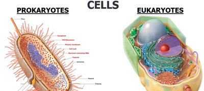
Cells are the fundamental units of life, and in microbiology, they are classified into two main types: prokaryotic and eukaryotic cells. Understanding their structural and functional differences is essential for studying microorganisms.
Classification of Organisms by Cell Type
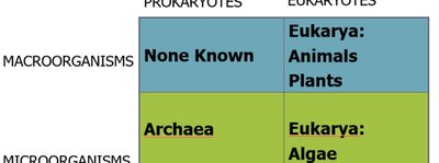
Prokaryotes: Include Bacteria and Archaea. No known prokaryotic macroorganisms.
Eukaryotes: Include Animals, Plants, Algae, Fungi, and Protozoa.
Prokaryotes | Eukaryotes | |
|---|---|---|
Macroorganisms | None Known | Eukarya: Animals, Plants |
Microorganisms | Archaea, Bacteria | Eukarya: Algae, Fungi, Protozoa |
Structural Differences Between Prokaryotic and Eukaryotic Cells
Prokaryotic and eukaryotic cells differ in their internal organization, genetic material, and cellular components.
Prokaryotes: DNA is not enclosed in a nuclear membrane; usually a single circular chromosome; no membrane-bound organelles; cell wall is complex if present; DNA not associated with histones.
Eukaryotes: DNA is enclosed in a nuclear membrane; multiple chromosomes; contains membrane-bound organelles (e.g., Golgi complex, mitochondria, lysosomes); cell wall is simple if present; DNA associated with histones and non-histones.
Terminology
Prokaryote: From Greek for "prenucleus" (no true nucleus).
Eukaryote: From Greek for "true nucleus" (contains a nucleus).

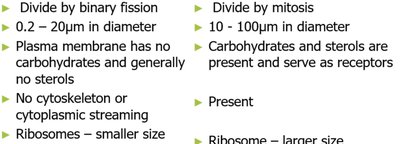
Further Differences in Cell Structure and Function
Prokaryotes: Divide by binary fission; size ranges from 0.2–20 μm; plasma membrane lacks carbohydrates and sterols; no cytoskeleton or cytoplasmic streaming; ribosomes are smaller; reproduction is asexual (may transfer DNA fragments).
Eukaryotes: Divide by mitosis; size ranges from 10–100 μm; carbohydrates and sterols present in plasma membrane (serve as receptors); cytoskeleton present; ribosomes are larger; sexual reproduction involves meiosis.
The Prokaryotes
Prokaryotes include Bacteria and Archaea. Some bacteria, such as Cyanobacteria, are photosynthetic. Bacterial species are differentiated by morphology, chemical composition, nutritional requirements, biochemical activities, and energy sources.
Morphology: Shape of the cell
Chemical composition: Detected by staining
Nutritional requirements
Biochemical activities
Source of energy: Sunlight or chemicals

Basic Shapes of Bacteria
Bacteria exhibit several basic shapes, which are important for identification and classification.
Bacillus: Rod-shaped
Coccus: Spherical
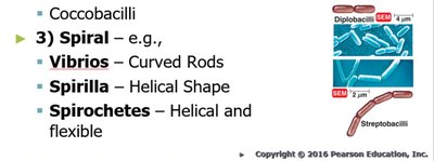
Spiral: Includes Spirillum, Vibrio, and Spirochete

Shapes of Bacterial Cells
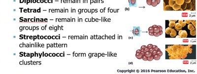
Bacterial cells can be arranged in various patterns depending on their division and grouping.
Coccus: Round, spherical, oval, or elongated; arrangements include single, diplococci (pairs), tetrad (groups of four), sarcinae (groups of eight), streptococci (chains), staphylococci (clusters).
Bacillus: Rod-shaped; arrangements include single, diplobacilli (pairs), streptobacilli (chains), coccobacilli (short rods).
Spiral: Includes Vibrios (curved rods), Spirilla (helical shape), Spirochetes (helical and flexible).
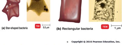
Unusual Bacterial Shapes
Star-shaped: Stella
Square: Haloarcula
Monomorphic and Pleomorphic Bacteria
The shape of a bacterium is determined by heredity. Most bacteria are monomorphic (maintain a single shape), but some are pleomorphic (can have more than one genetically controlled shape).
Monomorphic: Maintain a single shape; environmental factors may change their shape, making identification difficult.
Pleomorphic: Can have more than one shape; examples include Rhizobium and Corynebacterium.

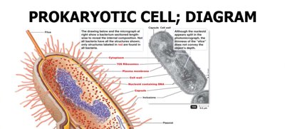
Prokaryotic Cell Diagram
The prokaryotic cell is characterized by its lack of a nucleus and membrane-bound organelles. Key structures include the plasma membrane, cell wall, capsule, cytoplasm, ribosomes, and flagella.
Structures External to the Cell Wall: Glycocalyx
The glycocalyx is a sugar coat found on the surface of many cells. In bacteria, it is a viscous, gelatinous polymer composed of polysaccharide and/or polypeptide. It can be organized as a capsule (firmly attached) or a slime layer (unorganized and loose).
Capsule: Organized and firmly attached glycocalyx
Slime layer: Unorganized and loosely attached glycocalyx

Additional info:
Capsules can protect bacteria from phagocytosis and aid in adherence to surfaces.
Slime layers facilitate motility and biofilm formation.
