 Back
BackFunctional Anatomy of Prokaryotic and Eukaryotic Cells: Cell Envelope & Biological Membranes
Study Guide - Smart Notes
 Tailored notes based on your materials, expanded with key definitions, examples, and context.
Cell Envelope & Biological Membranes
Cell Envelope: Structure and Components
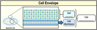
The cell envelope refers to all layers surrounding the cell, including membranes and cell walls. The composition of the cell envelope varies among different cell types, but all cells possess a cell membrane as a fundamental component. The cell envelope provides structural integrity and protection to the cell.
Cell membrane: The innermost layer, present in all cells.
Cell wall: Provides rigidity and protection, present in most prokaryotes.
Biological Membranes: Composition and Fluid Mosaic Model
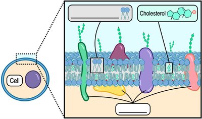
Biological membranes are primarily composed of phospholipids, which are amphipathic molecules. These membranes also contain embedded proteins and, in eukaryotes, cholesterol. The fluid mosaic model describes membranes as dynamic structures with proteins moving laterally within the lipid bilayer.
Phospholipid bilayer: Major structural framework.
Proteins: Integral and peripheral, perform various functions.
Cholesterol: Present in eukaryotic membranes, regulates fluidity.
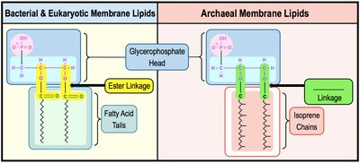
Phospholipid Structure
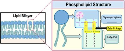
Phospholipids consist of a hydrophilic glycerophosphate head and two hydrophobic fatty acid tails. In bacteria and eukaryotes, these are connected by an ester linkage. The amphipathic nature allows the formation of bilayers, essential for membrane function.
Hydrophilic head: Glycerophosphate group.
Hydrophobic tails: Fatty acids.
Ester linkage: Connects head to tails in bacteria/eukaryotes.
Bacterial vs. Eukaryotic Cell Membranes
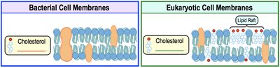
Bacterial membranes lack cholesterol, making them less rigid than eukaryotic membranes. Eukaryotic membranes contain cholesterol and lipid rafts, which are dense regions of cholesterol and proteins that move together, contributing to membrane fluidity and function.
Bacterial membranes: No cholesterol.
Eukaryotic membranes: Contain cholesterol and lipid rafts.
Archaeal Cell Membranes
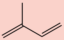
Archaeal membranes differ significantly from bacterial and eukaryotic membranes. Their hydrophobic tails are repeating isoprene units (5-carbon hydrocarbons), not fatty acids, and are connected to the glycerophosphate head by an ether linkage, which is more resistant to heat and chemical toxins.
Isoprene chains: Unique to archaea.
Ether linkage: Provides stability in extreme environments.
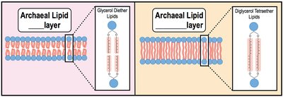
Types of Archaeal Membrane Lipids
Archaeal membrane lipids can form either bilayers or monolayers, depending on the lipid type. Monolayers, formed by diglycerol tetraether lipids, are more rigid and are found in thermophilic archaea, providing protection in extreme temperatures.
Bilayers: Glycerol diether lipids.
Monolayers: Diglycerol tetraether lipids, increase rigidity.
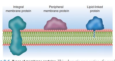
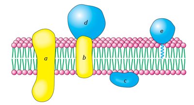
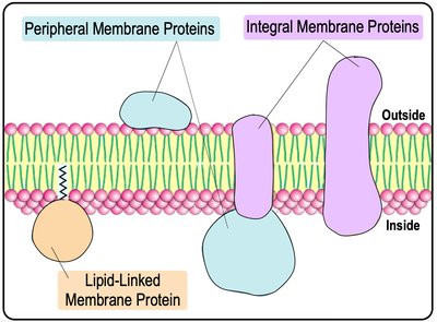
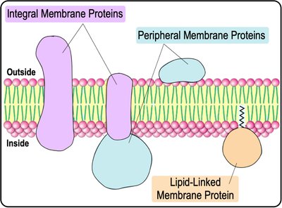
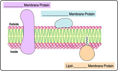
Types of Membrane Proteins
Classification of Membrane Proteins
Membrane-associated proteins are classified into three main types:
Integral proteins: Span the entire lipid bilayer, noncovalently integrated.
Peripheral proteins: Located on the periphery of the bilayer.
Lipid-anchored proteins: Covalently attached to lipid groups within the bilayer.
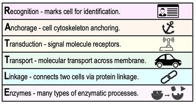
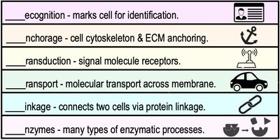
Functions of Membrane Proteins
Membrane proteins perform a wide variety of functions, including:
Recognition: Markers for cell identification.
Anchorage: Attachment to the extracellular matrix (ECM) and cytoskeleton.
Transduction: Receptors for signal-transduction pathways.
Transport: Molecular transport across the membrane.
Linkage: Connects two cells via protein linkages.
Enzymes: Catalyze various enzymatic processes.
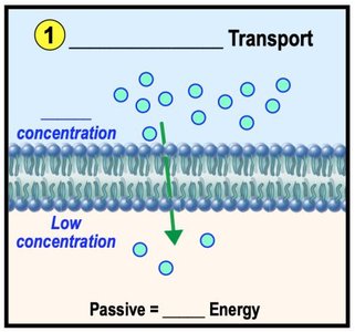
Concentration Gradients & Diffusion
Concentration Gradients
A concentration gradient is the difference in concentration of a substance between two areas. Molecules move down their gradient (from high to low concentration) passively, or up their gradient (from low to high concentration) actively, which requires energy.
Down gradient: Passive movement, no energy required.
Up gradient: Active movement, energy required.
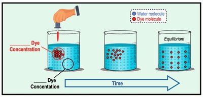
Diffusion
Diffusion is the movement of a substance from an area of high concentration to an area of low concentration. Molecules naturally diffuse down their concentration gradients.
Passive process: No energy required.
Equilibrium: Achieved when concentrations are equal.
Membrane Transport
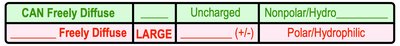
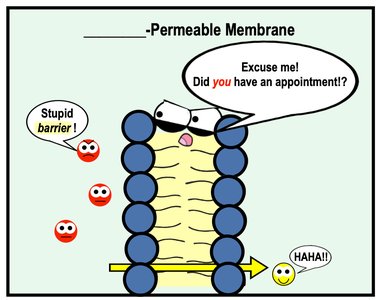
Selective Permeability
Biological membranes are selectively permeable, meaning they regulate what crosses the membrane. Some molecules can freely diffuse across the membrane without protein facilitation, while others require transport proteins.
Freely diffusing molecules: Small, uncharged, nonpolar (e.g., O2, CO2).
Require facilitation: Large, charged, or polar molecules.
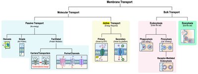
Overview of Membrane Transport Mechanisms
Membrane transport is divided into molecular and bulk transport. Molecular transport includes passive (no energy) and active (energy required) mechanisms, while bulk transport involves endocytosis and exocytosis for large molecules.
Passive transport: Simple diffusion, facilitated diffusion, osmosis.
Active transport: Primary and secondary active transport.
Bulk transport: Endocytosis (phagocytosis, pinocytosis, receptor-mediated), exocytosis.
Passive vs. Active Transport
There are two general types of molecular transport across biological membranes:
Passive transport: Moves molecules from high to low concentration, does not require energy.
Active transport: Moves molecules from low to high concentration, requires energy (usually ATP).
Classes of Membrane Transport Proteins
Transport proteins are classified by their operation:
Uniporters: Transport one molecule at a time in one direction.
Symporters: Cotransport two or more molecules in the same direction.
Antiporters: Cotransport two or more molecules in opposite directions.
Osmosis
Osmosis and Tonicity
Osmosis is the passive diffusion of water across a semi-permeable membrane. The direction of water flow depends on the relative concentration of solutes (tonicity) in the solutions.
Hypotonic: Lower solute concentration.
Isotonic: Equal solute concentration.
Hypertonic: Higher solute concentration.
Simple & Facilitated Diffusion
Simple Diffusion
Simple diffusion is the direct movement of small, uncharged molecules through the cell membrane without energy or protein assistance.
Examples: O2, CO2, N2.
Facilitated Diffusion
Facilitated diffusion is the passive movement of charged or polar molecules across the membrane, assisted by transport proteins such as channels or carriers.
Examples: Ions (Na+, Ca2+), glucose.
Transport Proteins in Facilitated Diffusion
Two main types of transport proteins are involved:
Porins/Channels: Form membrane-spanning pores, e.g., aquaporins for water.
Transporters/Carriers: Undergo conformational changes to move molecules.
Active Transport
Primary Active Transport
Primary active transport is directly driven by ATP hydrolysis, moving molecules against their concentration gradient. The sodium-potassium pump (Na+/K+ antiporter) is a classic example.
Na+/K+ pump: Exports 3 Na+ ions, imports 2 K+ ions.
Secondary Active Transport
Secondary active transport uses the concentration gradient established by primary active transport to power the movement of other molecules. For example, the Na+-glucose cotransporter uses the Na+ gradient to import glucose against its gradient.
ABC Transporters
ATP-Binding Cassette (ABC) transporters are integral membrane proteins with two trans-membrane domains and two nucleotide-binding domains. They pump substances across membranes against their gradient and are responsible for multidrug resistance in bacteria and humans.
Group Translocation
Group translocation is a special type of transport where a molecule is chemically modified as it enters the cell, such as phosphorylation in the E. coli phosphotransferase system (PTS). This process is exclusive to prokaryotes.
Endocytosis & Exocytosis
Endocytosis
Endocytosis is the process by which cells engulf large biomolecules via the cell membrane, forming vesicles. Types include phagocytosis (cell eating), pinocytosis (cell drinking), and receptor-mediated endocytosis (selective uptake).
Exocytosis
Exocytosis is the process by which vesicles fuse with the cell membrane to release their contents to the extracellular space. This is important for secretion of hormones, neurotransmitters, and digestive enzymes.
Prokaryotic & Eukaryotic Cells
Prokaryotic Cells
Prokaryotic cells lack a nucleus and membrane-bound organelles. Their DNA is circular and located in the nucleoid region. They have small (70S) ribosomes and divide by binary fission.
Eukaryotic Cells
Eukaryotic cells have a nucleus and membrane-bound organelles. Their DNA is linear and enclosed within the nucleus. They have large (80S) ribosomes and divide by mitosis and cytokinesis.
Binary Fission
Binary fission is the process by which prokaryotes replicate. The cell elongates, replicates DNA, forms a septum, and divides into two identical daughter cells. The time required for binary fission is called the generation time.
Generation Times
Generation time (doubling time) is the time it takes for a microbial population to double in number. The equation for calculating cell number is:
Where is the final number of cells, is the initial number, and is the number of generations.
Bacterial Cell Morphology & Arrangements
Cell Morphology
Bacterial cells exhibit various shapes: cocci (spherical), bacilli (rod-shaped), and spiral (spirillum, spirochete, vibrio). Cell arrangement refers to the grouping of cells after division, such as chains (strepto-), clusters (staphylo-), and pairs (diplo-).
Overview of Prokaryotic Cell Structures
Cell Wall
The cell wall is a semi-rigid structural layer outside the cell membrane, providing protection against osmotic pressure. The main component is peptidoglycan, a mesh-like polysaccharide and protein mix.
Peptidoglycan Structure
Peptidoglycan consists of repeating units of N-acetylglucosamine (NAG) and N-acetylmuramic acid (NAM) linked by β-(1,4) glycosidic bonds. Tetrapeptide chains attached to NAM molecules form peptide interbridges, especially in gram-positive bacteria.
Gram-Positive vs. Gram-Negative Cell Walls
Gram-positive bacteria have a thick peptidoglycan layer and teichoic acids. Gram-negative bacteria have a thin peptidoglycan layer, an outer membrane with lipopolysaccharides (LPS), and lipoproteins anchoring the membrane.
Glycocalyx: Capsules & Slime Layers
The glycocalyx is a polysaccharide layer surrounding the cell, promoting attachment and protection. Capsules are organized and tightly anchored, while slime layers are unorganized and easily removable.
Biofilms
Biofilms are groups of cells encased in a polysaccharide matrix (EPS) attached to surfaces, containing various microbes and polymers.
Pili, Fimbriae, and Hami
Pili are filamentous proteins involved in motility and DNA transfer (conjugation). Fimbriae are shorter and promote biofilm formation. Hami are unique to archaea, allowing attachment within microbial communities.
Flagella
Flagella are long filamentous proteins driving cell motility. Distribution patterns (monotrichous, peritrichous, amphitrichous, lophotrichous) help identify bacteria. The basal body structure differs between gram-positive and gram-negative cells.
Flagellar Movement & Chemotaxis
Flagellar motility involves runs and tumbles, powered by proton motive force (PMF). Chemotaxis is movement toward attractants or away from repellents, with cells adjusting run length based on concentration changes.
Prokaryotic Ribosomes
Prokaryotic ribosomes (70S) consist of 50S and 30S subunits, made of proteins and rRNA. Archaeal ribosomes differ in rRNA sequence and protein content, making them less susceptible to antibiotics targeting bacterial ribosomes.
Bacterial Plasmids
Plasmids are small, circular DNA molecules replicated independently of the chromosome, often carrying antibiotic resistance genes. Episomes are plasmids that integrate into the chromosome.
Cell Inclusions
Inclusions are cytoplasmic aggregates for storage (e.g., gas vesicles for buoyancy, magnetosomes for iron).
Endospores
Endospores are dormant, heat-resistant cells produced by Bacillus and Clostridium species for survival under adverse conditions. Sporulation is the process of endospore formation; germination is the return to vegetative state.
Eukaryotic Cell Structures
Eukaryotic Organelles
Eukaryotic cells contain membrane-bound organelles, including the nucleus, endoplasmic reticulum (ER), Golgi apparatus, lysosomes, peroxisomes, central vacuole (plants), mitochondria, and chloroplasts.
Endomembrane System
The endomembrane system includes interconnected organelles for protein secretion and cellular digestion. Proteins are synthesized in the nucleus, processed in the ER, modified in the Golgi, and secreted via vesicles.
Mitochondria & Chloroplasts
Mitochondria generate ATP via cellular respiration; chloroplasts perform photosynthesis. Both have their own DNA and ribosomes, supporting the endosymbiotic theory.
Cytoskeleton
The cytoskeleton is a network of proteins providing structure, motility, and transport. Components include microfilaments (actin), intermediate filaments, and microtubules (tubulin).
Eukaryotic Cilia & Flagella
Cilia and flagella are microtubule-based structures for cell movement, arranged in a 9+2 pattern. Cilia move like oars; flagella move in a whip-like motion.
Cell Junctions
Cell junctions link adjacent cells: tight junctions (impermeable barrier), anchoring junctions (desmosomes), gap junctions (exchange of molecules), and plasmodesmata (plant cells).
