 Back
BackChapter 4 - Functional Anatomy of Prokaryotic and Eukaryotic Cells: Study Notes
Study Guide - Smart Notes
 Tailored notes based on your materials, expanded with key definitions, examples, and context.
Functional Anatomy of Prokaryotic and Eukaryotic Cells
Overview: Comparing Prokaryotic and Eukaryotic Cells
This section introduces the fundamental differences between prokaryotic and eukaryotic cells, which are central to understanding microbial life. The structural and genetic distinctions between these cell types underpin their classification and biological functions.
Prokaryotes: Derived from Greek for 'prenucleus', these cells lack a membrane-bound nucleus. They possess a single circular chromosome, no histones, and no organelles. Bacteria have peptidoglycan cell walls, while archaea have pseudomurein cell walls. Prokaryotes divide by binary fission.
Eukaryotes: Derived from Greek for 'true nucleus', these cells have paired chromosomes within a nuclear membrane, histones, and organelles. Their cell walls, when present, are made of polysaccharides. Eukaryotes divide by mitosis.
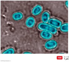
The Size, Shape, and Arrangement of Bacterial Cells
Bacterial morphology is diverse, with size, shape, and arrangement serving as key identifiers in microbiology. Most bacteria are monomorphic, but some exhibit pleomorphism.
Average size: 0.2 to 2.0 μm in diameter, 2 to 8 μm in length.
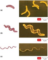
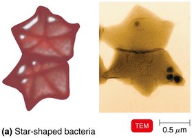
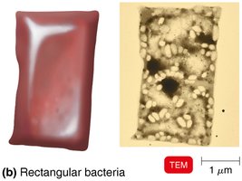
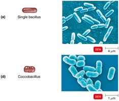
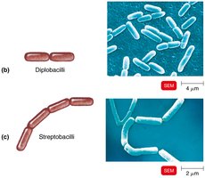
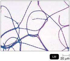
Shapes: Bacillus (rod-shaped), coccus (spherical), spiral (vibrio, spirillum, spirochete), star-shaped, and rectangular.
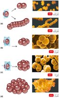
Arrangements: Pairs (diplococci, diplobacilli), clusters (staphylococci), chains (streptococci, streptobacilli), tetrads (groups of four), sarcinae (cubelike groups of eight).
Prokaryotic Cell Structure
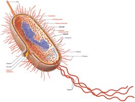
Prokaryotic cells are characterized by a simple internal structure, lacking membrane-bound organelles. Their cell envelope and appendages play crucial roles in protection, motility, and interaction with the environment.
Cell wall: Provides structural support and prevents osmotic lysis.
Plasma membrane: Regulates transport of substances.
Ribosomes: Sites of protein synthesis (measured in svedberg units, S).
Nucleoid: Region containing the circular chromosome.
External structures: Glycocalyx, flagella, fimbriae, pili.
Glycocalyx
The glycocalyx is an external, viscous, gelatinous layer composed of polysaccharides and/or polypeptides. It exists in two forms: capsule (organized, firmly attached) and slime layer (unorganized, loose).
Virulence factor: Capsules prevent phagocytosis, aiding pathogenicity.
Biofilm formation: Extracellular polymeric substances help bacteria adhere and form biofilms.
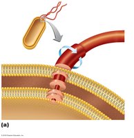
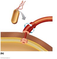
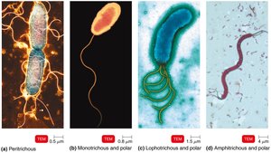
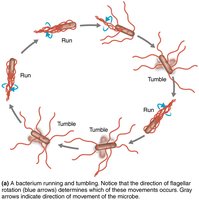
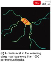
Flagella
Flagella are filamentous appendages that propel bacteria. They are composed of flagellin and consist of three parts: filament, hook, and basal body. The basal body anchors the flagellum to the cell wall and membrane.
Gram-negative bacteria: Basal body has 4 rings.
Gram-positive bacteria: Basal body has 2 rings.
Motility: Flagella rotate to produce 'run' and 'tumble' movements, enabling taxis (movement toward or away from stimuli).
H antigens: Flagella proteins serve as antigens for serovar identification (e.g., Escherichia coli O157:H7).
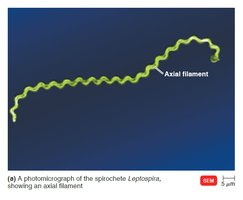
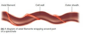
Axial Filaments
Axial filaments, also known as endoflagella, are specialized structures found in spirochetes. They are anchored at one end and rotate, causing the cell to move in a corkscrew fashion.
Function: Enables motility in viscous environments.
Location: Wrap around the cell beneath the outer sheath.
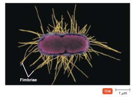
Fimbriae and Pili
Fimbriae and pili are hairlike appendages found on prokaryotic cells. Fimbriae are primarily involved in attachment, while pili are involved in motility and genetic exchange.
Fimbriae: Allow bacteria to adhere to surfaces and each other, facilitating colonization.
Pili: Involved in twitching motility and conjugation (DNA transfer between cells).
The Cell Wall: Structure and Function
The bacterial cell wall is essential for maintaining cell shape, protecting against osmotic lysis, and contributing to pathogenicity. Its composition varies between gram-positive and gram-negative bacteria.
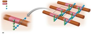
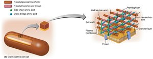
Peptidoglycan: A polymer of repeating disaccharides (N-acetylglucosamine [NAG] and N-acetylmuramic acid [NAM]) linked by polypeptides.
Gram-positive cell walls: Thick peptidoglycan, teichoic acids (lipoteichoic and wall teichoic acids), high susceptibility to penicillin.
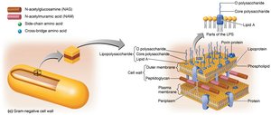
Gram-negative cell walls: Thin peptidoglycan, outer membrane, periplasmic space, low susceptibility to penicillin.
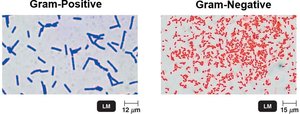
Gram Stain Mechanism
The Gram stain is a differential staining technique that distinguishes bacteria based on cell wall structure. It is a fundamental tool in microbiology for classification and diagnosis.
Gram-positive: Alcohol dehydrates peptidoglycan, trapping crystal violet-iodine (CV-I) complexes; cells appear purple.
Gram-negative: Alcohol dissolves the outer membrane and creates holes in peptidoglycan, allowing CV-I to wash out; cells are colorless until counterstained with safranin (appear red/pink).
Summary Table: Gram-Positive vs. Gram-Negative Bacteria
The following table summarizes key differences between gram-positive and gram-negative bacteria, including cell wall structure, susceptibility to antibiotics, and pathogenic features.
Feature | Gram-Positive | Gram-Negative |
|---|---|---|
Peptidoglycan Thickness | Thick | Thin |
Teichoic Acids | Present | Absent |
Outer Membrane | Absent | Present |
Susceptibility to Penicillin | High | Low |
Pathogenicity | Exotoxins | Endotoxins & Exotoxins |
Basal Body Rings (Flagella) | 2 | 4 |
Key Terms and Concepts
Peptidoglycan: Structural polymer in bacterial cell walls.
Teichoic acids: Polymers in gram-positive cell walls, contribute to antigenic specificity.
Lipopolysaccharide (LPS): Component of gram-negative outer membrane; includes O polysaccharide (antigen) and Lipid A (endotoxin).
Binary fission: Method of prokaryotic cell division.
Mitosis: Method of eukaryotic cell division.
Equations and Formulas
Peptidoglycan structure can be represented as:
Cross-linking occurs via peptide bonds between tetrapeptide side chains.
Additional info: Academic context was added to clarify the functions and significance of cell wall components, motility structures, and staining mechanisms. The summary table was inferred and expanded for completeness.
