 Back
Functional Anatomy of Prokaryotic and Eukaryotic Cells: Study Guide
Study Guide - Smart Notes
 Tailored notes based on your materials, expanded with key definitions, examples, and context.
Functional Anatomy of Prokaryotic and Eukaryotic Cells
Distinguishing Bacterial Genus vs. Shape
In microbiology, the term bacillus can refer to both a bacterial genus (Bacillus) and a rod-shaped morphology. To determine which is meant, context is key:
Genus: Capitalized and italicized (e.g., Bacillus anthracis).
Shape: Lowercase, not italicized, refers to rod-shaped bacteria.
Example: "Bacillus causes anthrax" refers to the genus; "bacillus-shaped bacteria" refers to morphology.
Endospore Formation in Bacteria
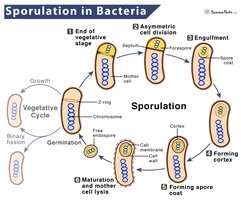
Endospore formation is a survival mechanism in certain bacteria, allowing them to withstand harsh conditions. The process involves several stages:
Sporulation: The process of endospore formation.
Initiation: Triggered by nutrient depletion or environmental stress.
Germination: Formation of a new cell from an endospore, triggered by favorable conditions.
Example: Bacillus and Clostridium species form endospores to survive extreme environments.
Structure and Function of Bacterial Cell Walls
Bacterial cell walls are essential for maintaining cell shape, protecting against osmotic lysis, and providing structural integrity. There are several types:
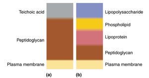
Gram-positive: Thick peptidoglycan layer, teichoic acids.
Gram-negative: Thin peptidoglycan layer, outer membrane with lipopolysaccharides.
Acid-fast: Contains mycolic acids, resistant to decolorization.
Mycoplasmas: Lack cell walls, have sterols in membrane.
Example: Gram staining distinguishes between gram-positive and gram-negative bacteria based on cell wall structure.
Comparison Table: Bacterial Cell Walls
Type | Main Features | Gram Stain | Drug Sensitivity |
|---|---|---|---|
Gram-positive | Thick peptidoglycan, teichoic acids | Purple | Sensitive to penicillin |
Gram-negative | Thin peptidoglycan, outer membrane, LPS | Pink | Resistant to penicillin |
Acid-fast | Mycolic acids, waxy layer | Variable | Resistant to many drugs |
Mycoplasmas | No cell wall, sterols in membrane | No stain | Resistant to cell wall-targeting drugs |
Gram Stain Mechanism and Drug Effects
The Gram stain differentiates bacteria based on cell wall structure:
Gram-positive: Retain crystal violet due to thick peptidoglycan.
Gram-negative: Lose crystal violet, counterstained pink.
Penicillin: Inhibits peptidoglycan synthesis, effective against gram-positive.
Gram-negative resistance: Outer membrane blocks penicillin entry.
Example: Penicillin is ineffective against most gram-negative bacteria due to their outer membrane.
Peptidoglycan Structure and Synthesis
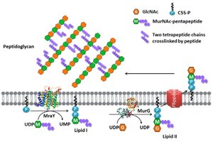
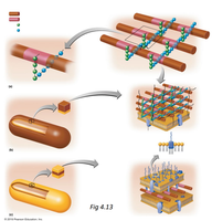
Peptidoglycan is a polymer of sugars and amino acids forming a mesh-like layer outside the plasma membrane. Its synthesis is a target for antibiotics:
Structure: Alternating N-acetylglucosamine (GlcNAc) and N-acetylmuramic acid (MurNAc) with peptide crosslinks.
Synthesis: Enzymes assemble peptidoglycan precursors and transport them across the membrane.
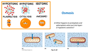
Osmosis and Effects on Bacterial Cells
Osmosis is the movement of water across a semipermeable membrane. Bacterial cells respond differently to various solutions:
Isotonic: No net water movement; cell remains stable.
Hypotonic: Water enters cell; risk of lysis if cell wall is damaged.
Hypertonic: Water leaves cell; plasmolysis occurs.
Protoplasts and spheroplasts (cells lacking cell wall) are highly sensitive to osmotic changes:
Hypertonic solution: Cell shrinks (plasmolysis).
Hypotonic solution: Cell swells and may lyse (cytolysis).
Fimbriae and Other Bacterial Structures
Fimbriae are proteinaceous appendages found on many gram-negative bacteria:
Function: Attachment to surfaces and host cells.
Composition: Made of pilin protein.
Motility: Not used for motility (unlike flagella).
Example: Escherichia coli uses fimbriae to adhere to urinary tract epithelium.
Clinical Applications: Anaerobic Bacteria and Environmental Survival
Clostridium botulinum is a strict anaerobe, killed by oxygen. It survives on plants as endospores, which are resistant to environmental stress. Home-canned foods are a common source of botulism because improper canning fails to destroy endospores, allowing them to germinate in anaerobic conditions.
Endospores: Allow survival in oxygen-rich environments until anaerobic conditions are met.
Home-canning: Often lacks sufficient heat to destroy endospores.
Environmental Microbiology: Bacterial Contamination in Water
Bacteria such as Acidithiobacillus can enter household water through biofilms or pipe contamination. Structures like fimbriae and pili facilitate attachment to surfaces, enabling colonization of plumbing systems.
Biofilms: Communities of bacteria adhering to surfaces, protected by extracellular matrix.
Fimbriae: Aid in attachment and persistence in water systems.
Analysis and Application Questions
Epulopiscium fishelsoni: Initially classified as protozoan due to large size; reclassified as gram-positive bacterium based on cell wall structure, DNA, and ribosomal RNA analysis.
Active Transport of K+ in Hypertonic Solutions: Helps maintain osmotic balance and prevents plasmolysis by increasing internal solute concentration, requiring ATP.
Summary Table: Effects of Solutions on Bacterial Cells
Cell Type | Isotonic | Hypotonic | Hypertonic |
|---|---|---|---|
Intact cell | No change | Water enters, possible lysis | Water leaves, plasmolysis |
Protoplast/Spheroplast | No change | Water enters, lysis | Water leaves, shrinkage |
