 Back
BackFunctional Anatomy of Prokaryotic and Eukaryotic Cells: Study Notes
Study Guide - Smart Notes
 Tailored notes based on your materials, expanded with key definitions, examples, and context.
Functional Anatomy of Prokaryotic and Eukaryotic Cells
Comparing Prokaryotic and Eukaryotic Cells
Prokaryotic and eukaryotic cells differ fundamentally in their structure and organization. Understanding these differences is essential for microbiology, as it underpins the classification and physiology of microorganisms.
Prokaryotes: Cells lacking a true nucleus and membrane-bound organelles. Their genetic material is a single, circular DNA molecule not enclosed by a nuclear membrane. They typically have peptidoglycan cell walls and divide by binary fission.
Eukaryotes: Cells with a true nucleus surrounded by a nuclear membrane. Their DNA is linear and associated with histone proteins. Eukaryotes possess various organelles and, when present, their cell walls are made of polysaccharides. They divide by mitosis or meiosis.
The Size, Shape, and Arrangement of Bacterial Cells
Size and Morphology
Bacteria exhibit a variety of shapes and arrangements, which are important for identification and classification.
Average size: 0.2 to 2.0 µm in diameter and 2 to 8 µm in length.
Monomorphic: Most bacteria maintain a single shape.
Pleomorphic: Some bacteria can alter their shape in response to environmental conditions.
Bacillus: Rod-shaped
Coccus: Spherical
Spiral forms: Vibrio (comma-shaped), Spirillum (rigid spiral), Spirochete (flexible spiral)
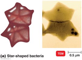
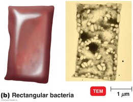
Other forms: Star-shaped, rectangular
Arrangements of Bacterial Cells
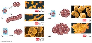
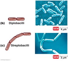
Bacteria can be found in characteristic arrangements due to their patterns of division and cellular adhesion.
Pairs (diplo-): Diplococci, diplobacilli
Chains (strepto-): Streptococci, streptobacilli
Clusters (staphylo-): Staphylococci
Groups of four: Tetrads
Cubelike groups of eight: Sarcinae
The Structure of a Prokaryotic Cell
Glycocalyx
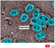
The glycocalyx is a general term for substances that surround bacterial cells. It plays a role in protection and adherence.
Capsule: Organized and firmly attached to the cell wall; protects against phagocytosis.
Slime layer: Unorganized and loosely attached; aids in adherence and biofilm formation.
Composition: Polysaccharide and/or polypeptide.
Flagella
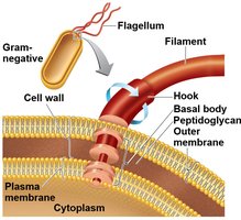
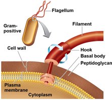
Flagella are long, whip-like appendages used for motility. Their arrangement and structure differ between Gram-positive and Gram-negative bacteria.
Structure: Composed of filament, hook, and basal body.
Function: Movement toward or away from stimuli (taxis), including chemotaxis and phototaxis.
Arrangements: Monotrichous (single), lophotrichous (tuft), amphitrichous (both poles), peritrichous (all over).
Axial Filaments
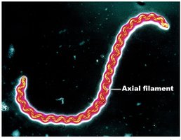
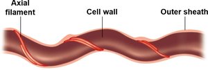
Axial filaments, or endoflagella, are found in spirochetes and enable corkscrew-like movement.
Location: Anchored at one end of the cell, wrapped around the cell body.
Function: Rotation causes the cell to move in a spiral motion.
Fimbriae and Pili
Fimbriae and pili are hairlike appendages on the surface of many bacteria, involved in attachment and genetic exchange.
Fimbriae: Numerous, short, and used for attachment to surfaces.
Pili: Longer, fewer (1-2 per cell), involved in motility and DNA transfer (conjugation).
The Cell Wall
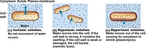
The bacterial cell wall is a semi-rigid structure that maintains cell shape and prevents osmotic lysis. It is a major target for antibiotics.
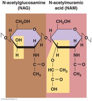
Composition: Peptidoglycan, a polymer of N-acetylglucosamine (NAG) and N-acetylmuramic acid (NAM) linked by polypeptides.
Function: Provides structural support and contributes to pathogenicity in some species.
Gram-Positive vs. Gram-Negative Cell Walls
Bacteria are classified as Gram-positive or Gram-negative based on their cell wall structure and response to Gram staining.
Gram-Positive: Thick peptidoglycan layers, teichoic acids, no outer membrane.
Gram-Negative: Thin peptidoglycan, outer membrane with lipopolysaccharides (LPS), lipoproteins, and phospholipids. The lipid A portion of LPS is an endotoxin.
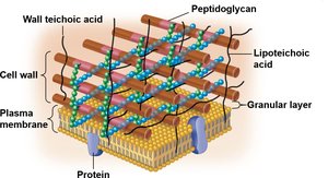
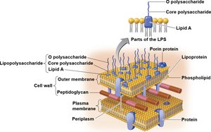
Table: Characteristics of Gram-Positive and Gram-Negative Bacteria
Feature | Gram-Positive | Gram-Negative |
|---|---|---|
Peptidoglycan | Thick | Thin |
Teichoic acids | Present | Absent |
Outer membrane | Absent | Present |
Lipopolysaccharide (LPS) | Absent | Present |
Sensitivity to penicillin | High | Low |
Gram stain color | Purple | Pink/red |
Atypical Cell Walls
Some bacteria, such as mycoplasmas, lack cell walls entirely and instead have sterols in their plasma membranes for stability.
Damage to the Cell Wall
Antibiotics like penicillin and enzymes like lysozyme target the bacterial cell wall, making them effective against Gram-positive bacteria. Gram-negative bacteria are more resistant due to their outer membrane.
The Plasma (Cytoplasmic) Membrane
Structure and Function
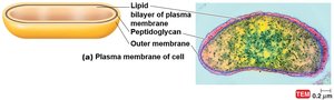
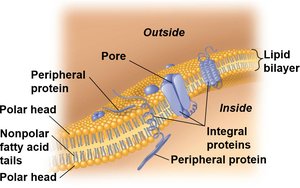
The plasma membrane is a phospholipid bilayer with embedded proteins, following the fluid mosaic model. It is selectively permeable and involved in energy production and transport.
Damage: Alcohols, detergents, and some antibiotics can disrupt the membrane, causing cell death.
Movement of Materials Across Membranes
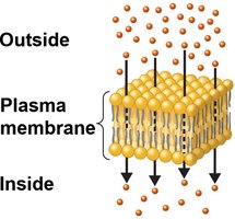
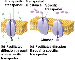
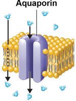
Transport across the membrane can be passive (no energy required) or active (requires energy).
Passive processes: Simple diffusion, facilitated diffusion, osmosis.
Active processes: Active transport, group translocation (unique to prokaryotes).
Internal Structures of Prokaryotic Cells
Cytoplasm
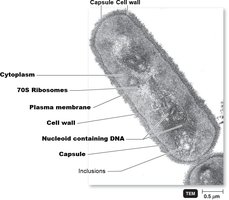
The cytoplasm is the substance inside the plasma membrane, consisting mostly of water, proteins, carbohydrates, lipids, and ions. It contains the cytoskeleton for cell shape.
Nucleoid
The nucleoid contains the bacterial chromosome (single, circular DNA) and may also contain plasmids, which are small, circular DNA molecules carrying non-essential genes.
Ribosomes
Ribosomes are the sites of protein synthesis. Prokaryotic ribosomes are 70S, making them susceptible to certain antibiotics that do not affect eukaryotic 80S ribosomes.
Inclusions
Inclusions are reserve deposits of nutrients and other substances, such as phosphate (metachromatic granules), glycogen, lipids, and sulfur.
Endospores
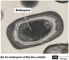
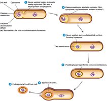
Endospores are highly resistant, dormant structures formed by some Gram-positive bacteria in response to adverse conditions. They can survive extreme heat, desiccation, chemicals, and radiation.
Sporulation: Formation of endospores.
Germination: Return to vegetative state when conditions improve.
Eukaryotic Cells
General Features
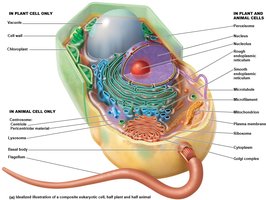
Eukaryotic cells are larger and more complex than prokaryotic cells. They contain a nucleus and various membrane-bound organelles.
Eukaryotic Cell Wall and Glycocalyx
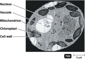
Cell walls are present in plants, fungi, and some algae, composed of carbohydrates such as cellulose, chitin, glucan, and mannan. Animal cells lack cell walls but may have a glycocalyx for protection and cell recognition.
The Plasma (Cytoplasmic) Membrane
Similar to prokaryotes but contains sterols and carbohydrates. Eukaryotic membranes can perform endocytosis (phagocytosis and pinocytosis).
Cytoplasm and Nucleus
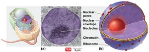
The cytoplasm contains a cytoskeleton for shape and support. The nucleus is surrounded by a nuclear envelope and contains DNA organized into chromosomes.
Ribosomes
Eukaryotic ribosomes are 80S (larger than prokaryotic 70S), found free in the cytoplasm or bound to the endoplasmic reticulum. Mitochondria and chloroplasts contain 70S ribosomes.
Endomembrane System
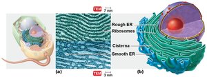
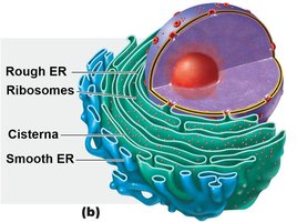
Endoplasmic Reticulum (ER): Rough ER (with ribosomes) synthesizes proteins; smooth ER synthesizes lipids and hormones.
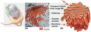
Golgi Complex: Modifies, sorts, and packages proteins and lipids for secretion or delivery to other organelles.
Mitochondria and Chloroplasts
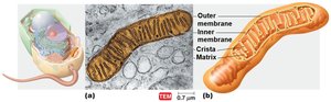
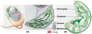
Mitochondria are the site of ATP production and contain their own DNA and 70S ribosomes. Chloroplasts, found in plants and algae, are the site of photosynthesis.
Other Organelles
Lysosomes: Contain digestive enzymes for breaking down waste.
Vacuoles: Storage organelles, especially prominent in plant cells.
