 Back
Functional Anatomy of Prokaryotic and Eukaryotic Cells
Study Guide - Smart Notes
 Tailored notes based on your materials, expanded with key definitions, examples, and context.
Functional Anatomy of Prokaryotic and Eukaryotic Cells
Overview
This chapter explores the structural and functional differences between prokaryotic and eukaryotic cells, focusing on bacterial morphology, anatomy, and the mechanisms by which substances move across cellular membranes. Understanding these differences is fundamental to microbiology, as it underpins microbial classification, physiology, and the basis for many antimicrobial strategies.
Prokaryotic vs. Eukaryotic Cells
Key Differences
Prokaryotic Cells: DNA is not enclosed in a membrane, lack membrane-bound organelles, contain peptidoglycan in their cell walls, and divide by binary fission. Their DNA is 'naked' (not associated with histones).
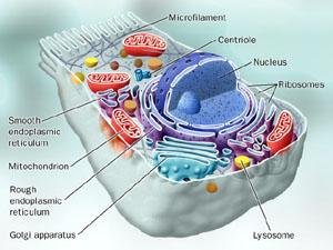
Eukaryotic Cells: Possess a true nucleus with DNA associated with histones, contain membrane-bound organelles (e.g., mitochondria, ER, Golgi apparatus), have simple cell walls when present, and divide by mitosis.
Size, Shape, and Arrangement of Bacterial Cells
Bacterial Morphology
Size: Typically 0.2–2.0 μm in diameter and 2–8 μm in length.
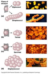
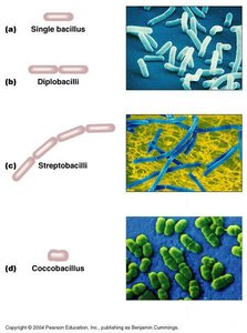
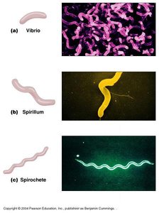
Shapes: Cocci (spherical), Bacilli (rod-shaped), Spirals (vibrio, spirillum, spirochete).
Arrangements: Determined by planes of division and whether cells remain attached after division (e.g., diplo-, strepto-, staphylo-).
Bacterial Anatomy
Major Structures
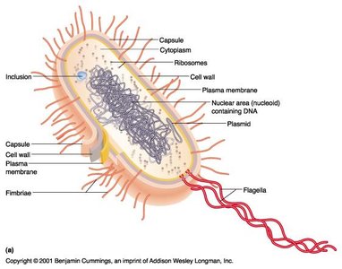
Glycocalyx: A polysaccharide/polypeptide layer outside the cell wall; can be a capsule (firmly attached) or slime layer (loosely attached). Functions in protection from phagocytosis, attachment, and environmental resistance.
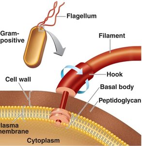
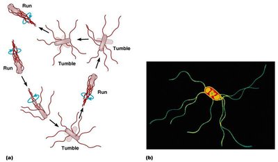
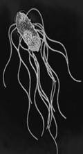
Flagella: Long, filamentous appendages for motility, composed of flagellin (filament), hook, and basal body. Enable taxis (movement toward/away from stimuli).
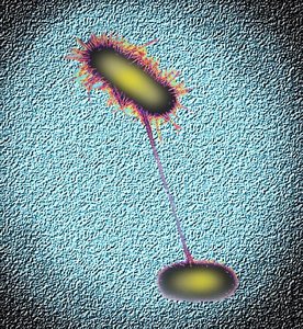
Fimbriae and Pili: Fimbriae are short, hair-like structures for attachment; pili are longer and involved in DNA transfer (conjugation) and motility.
Cell Wall: Provides shape and protection; composed mainly of peptidoglycan in bacteria.
Cytoplasm: Gel-like substance inside the plasma membrane containing water, proteins, DNA, ribosomes, and inclusions.
Nucleoid: Region containing the bacterial chromosome (circular, double-stranded DNA) and sometimes plasmids.
Ribosomes: Sites of protein synthesis; prokaryotic ribosomes are 70S (smaller than eukaryotic 80S).
Inclusions: Reserve deposits of nutrients and other substances.
Endospores: Highly resistant, dormant structures formed by some Gram-positive bacteria under stress.
Flagella and Motility
Structure and Function
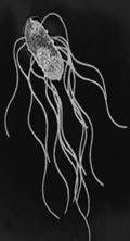
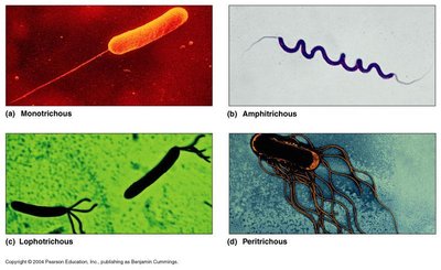
Flagella: Enable motility; composed of filament, hook, and basal body. The arrangement of flagella can be monotrichous (single), lophotrichous (tuft at one end), amphitrichous (one at each end), or peritrichous (all over the cell).
Motility: Bacteria move by "runs" (straight movement) and "tumbles" (random changes in direction), often in response to chemical (chemotaxis) or light (phototaxis) stimuli.
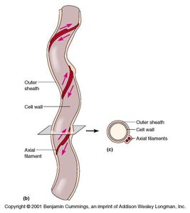
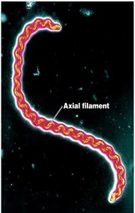
Axial Filaments (Endoflagella)
Found in spirochetes; wrap around the cell, anchored at one end, causing a corkscrew motion that enables movement through viscous environments (e.g., body fluids).
Fimbriae and Pili
Structure and Function
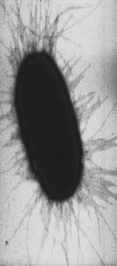
Fimbriae: Short, numerous, used for attachment to surfaces (important in pathogenesis).
Pili: Longer, usually 1–2 per cell; involved in DNA transfer (conjugation) and sometimes motility.
Bacterial Cell Wall
Composition and Function
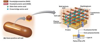
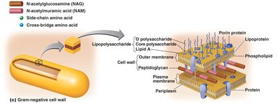
Peptidoglycan: Main component; consists of repeating disaccharides (N-acetylglucosamine and N-acetylmuramic acid) cross-linked by peptides.
Function: Maintains shape, prevents osmotic lysis, and provides protection.
Antibiotic Target: Penicillin inhibits peptidoglycan synthesis, leading to cell lysis.

Gram-Positive vs. Gram-Negative Cell Walls
Gram-Positive: Thick peptidoglycan layer, teichoic acids (regulate cation movement, cell growth).
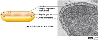
Gram-Negative: Thin peptidoglycan layer, outer membrane with lipopolysaccharides (LPS), lipoproteins, and phospholipids. The outer membrane acts as a barrier to certain antibiotics and contains lipid A (endotoxin).
Atypical Cell Walls
Mycoplasma: Lack cell walls; smallest bacteria.
Archaea: Contain pseudopeptidoglycan or other variants.
Mycobacterium: Contain mycolic acid (waxy lipid) instead of peptidoglycan.
Plasma Membrane
Structure and Function
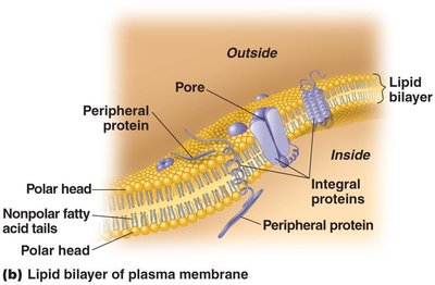
Phospholipid Bilayer: Fluid mosaic model; selectively permeable barrier.
Proteins: Integral and peripheral proteins serve as transporters, enzymes, or receptors.
Antimicrobial Target: Some antibiotics (e.g., polymyxins) disrupt the plasma membrane.
Movement of Materials Across Membranes
Passive Transport
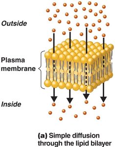
Simple Diffusion: Movement of molecules from high to low concentration without energy input.
Facilitated Diffusion: Utilizes carrier proteins (permeases) to transport substances down their concentration gradient.
Osmosis: Movement of water across a selectively permeable membrane. Types of solutions:
Isotonic: Equal solute concentration inside and outside the cell.
Hypotonic: Lower solute concentration outside; water enters, cell may lyse.
Hypertonic: Higher solute concentration outside; water leaves, cell shrinks (plasmolysis).

Active Transport
Requires energy (ATP) to move substances against their concentration gradient, often using transporter proteins.
Group Translocation: Substance is chemically modified during transport (e.g., glucose uptake in bacteria).
Cytoplasm
Composition and Function
80% water, contains proteins, carbohydrates, lipids, ions, DNA, ribosomes, and inclusions.
Site of most cellular processes.
Nuclear Area (Nucleoid) and Plasmids
Genetic Material
Nucleoid: Contains the bacterial chromosome (circular, double-stranded DNA), not surrounded by a membrane.
Plasmids: Small, circular DNA molecules; carry genes for antibiotic resistance, toxin production, and can be transferred between cells.
Ribosomes
Protein Synthesis
Composed of rRNA and protein; prokaryotic ribosomes are 70S, eukaryotic are 80S.
Target for many antibiotics (e.g., streptomycin, tetracycline).
Inclusions
Storage Granules
Reserve deposits for nutrients such as phosphate (volutin), glycogen, starch, lipids, sulfur, and gas vacuoles.
Endospores
Survival Structures
Formed by some Gram-positive bacteria (e.g., Bacillus, Clostridium) in response to adverse conditions.
Highly resistant to heat, desiccation, chemicals; important in food safety and sterilization.
Endospore formation is called sporulation; return to active state is germination.
Summary Table: Comparison of Prokaryotic and Eukaryotic Cells
Feature | Prokaryotic Cells | Eukaryotic Cells |
|---|---|---|
Nucleus | No true nucleus | True nucleus present |
DNA | Circular, naked, no histones | Linear, associated with histones |
Organelles | Absent | Present (mitochondria, ER, etc.) |
Cell Wall | Peptidoglycan (most) | Cellulose, chitin, or none |
Division | Binary fission | Mitosis/meiosis |
Ribosomes | 70S | 80S (cytoplasm), 70S (organelles) |
