 Back
Functional Anatomy of Prokaryotic and Eukaryotic Cells: Microbiology Study Notes
Study Guide - Smart Notes
 Tailored notes based on your materials, expanded with key definitions, examples, and context.
Functional Anatomy of Prokaryotic and Eukaryotic Cells
Overview: Similarities and Differences
Both prokaryotic and eukaryotic cells contain nucleic acids, proteins, lipids, and carbohydrates. They utilize similar chemical reactions for metabolism, protein synthesis, and energy storage. However, they differ significantly in structural organization and complexity.
Prokaryotic cells lack a membrane-bound nucleus and organelles.
Eukaryotic cells possess a nucleus and various membrane-bound organelles.
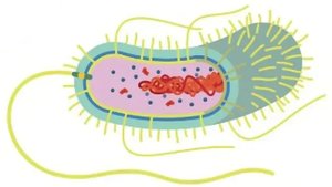
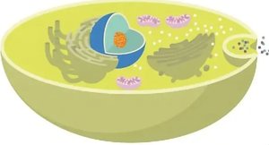
Prokaryotic and Eukaryotic Cell Structure
Prokaryotic cells are generally smaller (0.2–2.0 μm diameter, 2–8 μm length) and have shapes determined by genetics. Common shapes include bacillus (rod-shaped), coccus (spherical), and spiral (corkscrew).
Bacilli: Rod-shaped bacteria
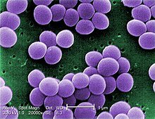
Cocci: Spherical bacteria
Spiral: Includes vibrio, spirillum, and spirochete forms
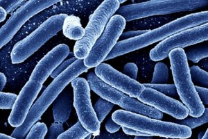
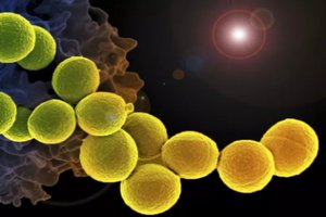
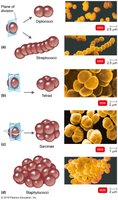
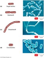
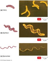
Bacterial Arrangements
Bacteria can be arranged in characteristic patterns:
Staphylo-: Grapelike clusters (cocci)
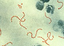
Strepto-: Chains (cocci or bacilli)
Prokaryotic Cell Anatomy
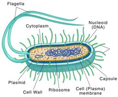
Prokaryotic cells contain several key structures:
Nucleoid: Region containing DNA
Plasmids: Small, circular DNA molecules
Ribosomes: Sites of protein synthesis
Cell wall: Provides shape and protection
Capsule: External protective layer
Flagella: Motility structures
Glycocalyx
Structure and Types
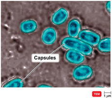
The glycocalyx is a viscous, gelatinous layer external to the cell wall, composed of polysaccharides and/or polypeptides. It exists in two forms:
Capsule: Neatly organized and firmly attached
Slime layer: Unorganized and loosely attached
Functions of Glycocalyx
Adherence: Helps bacteria attach to surfaces
Virulence: Capsules prevent phagocytosis, contributing to pathogenicity
Biofilm formation: EPS (extracellular polymeric substance) aids in cell attachment
Protection: Shields against dehydration and toxins
Nutrition: May serve as a nutrient source
Flagella and Other Appendages
Flagella Structure and Function
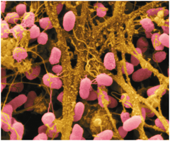
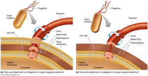
Flagella are long, whip-like appendages used for motility. Their structure differs between Gram-negative and Gram-positive bacteria.
Parts: Filament, hook, and basal body
Function: Movement via rotation
Flagella Arrangements
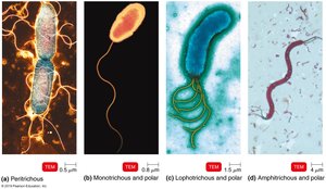
Peritrichous: Flagella all over the cell
Monotrichous: Single flagellum at one end
Lophotrichous: Multiple flagella at one end
Amphitrichous: Flagella at both ends
Flagella Movement
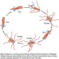
Bacteria move via taxis, responding to environmental stimuli (e.g., chemotaxis). Movement involves alternating "runs" and "tumbles" as flagella rotate.
Other Appendages: Axial Filaments, Fimbriae, Pili
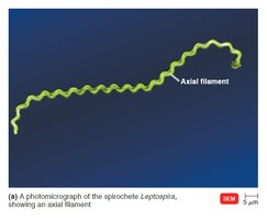
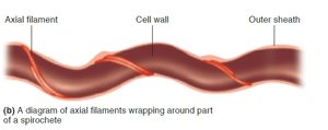
Axial filaments: Found in spirochetes, enable corkscrew movement
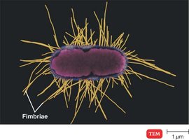
Fimbriae: Hairlike structures for attachment
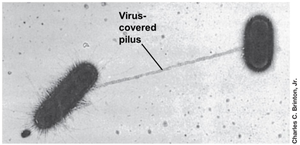
Pili: Used for motility and DNA transfer (conjugation)
Bacterial Cell Wall
Structure and Function
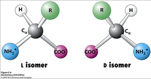
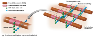
The bacterial cell wall prevents cell lysis due to osmotic pressure, maintains shape, and anchors flagella. It is primarily composed of peptidoglycan, which contains both D and L amino acids.
Peptidoglycan Structure
Peptidoglycan consists of a carbohydrate backbone (N-acetylglucosamine and N-acetylmuramic acid) linked by polypeptides.
Gram-Positive vs. Gram-Negative Cell Walls
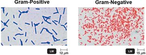
Bacteria are classified based on cell wall structure, which is revealed by the Gram stain.
Gram-positive: Thick peptidoglycan layers, teichoic acids
Gram-negative: Thin peptidoglycan layer, outer membrane with lipopolysaccharides, porins, and a gel-like periplasm
Plasma Membrane
Structure
The plasma membrane is a phospholipid bilayer separating the cell wall from the cytoplasm. It includes proteins and lipids, and follows the Fluid Mosaic Model.
Phosphate heads: Hydrophilic, face outward
Fatty acid tails: Hydrophobic, face inward
Functions
Selective permeability: Controls entry and exit of substances
Energy generation: Site of nutrient breakdown and ATP production
Movement of Substances Across Membranes
Passive Processes
Simple diffusion: Movement from high to low concentration, no ATP required
Facilitated diffusion: Movement via membrane proteins, no ATP required
Osmosis: Movement of water across membrane, no ATP required
Osmotic Pressure and Solutions
Isotonic: Equal solute concentration inside and outside cell
Hypotonic: Lower solute concentration outside cell
Hypertonic: Higher solute concentration outside cell
Active Processes
Active transport: Requires transporter protein and ATP; moves substances against concentration gradient
Group translocation: Requires transporter protein and PEP; substance is chemically altered during transport
Internal Structures of Prokaryotic Cells
Inclusions
Metachromatic granules: Store inorganic phosphate
Polysaccharide and lipid inclusions: Energy reserves
Sulfur granules: Energy source for sulfur bacteria
Carboxysomes: Contain enzymes for photosynthesis
Gas vacuoles: Provide buoyancy in aquatic bacteria
Magnetosomes: Contain iron oxide, act as magnets
Endospores
Endospores are highly durable, dehydrated cells formed by some Gram-positive bacteria. They are resistant to heat, dehydration, toxins, and radiation, and can survive for millions of years.
Eukaryotic Cell Structure
General Structure
Eukaryotic cells are larger and more complex, containing a nucleus and various organelles.
Eukaryotic Flagella and Cilia
Flagella and cilia are projections for motility or moving substances along the cell surface. Both contain microtubules arranged in a 9+2 array and move in a wave-like manner.
Flagella: Few and long (e.g., Euglena)
Cilia: Many and short (e.g., Tetrahymena)
Eukaryotic Cell Wall and Glycocalyx
Cell wall: Present in plants, algae, fungi; made of polysaccharides (cellulose, chitin, glucan, mannan)
Pellicle: Flexible protein covering in protozoans
Glycocalyx: Sticky carbohydrate layer covering plasma membrane; strengthens cell surface and aids attachment
Eukaryotic Plasma Membrane
Similar to prokaryotes, but contains sterols
Endocytosis: Plasma membrane engulfs extracellular material
Cytoplasm and Cytoskeleton
Cytoplasm: Substance inside plasma membrane and outside nucleus
Cytosol: Fluid portion
Cytoskeleton: Microtubules (tubulin), microfilaments (actin), intermediate filaments; maintain shape, anchor organelles, enable movement
Ribosomes
Sites of protein synthesis
Free in cytoplasm or bound to ER
Consist of large and small subunits
Nucleus
Contains DNA
Surrounded by nuclear envelope
Endoplasmic Reticulum (ER)
Rough ER: Protein processing and sorting; covered with ribosomes
Smooth ER: Synthesis of phospholipids, fats, steroids
Golgi Complex
Processes and sorts proteins from ER
Proteins leave via secretory vesicles, plasma membrane vesicles, or lysosomes
Lysosomes and Vacuoles
Lysosomes: Contain enzymes for molecule breakdown; digest bacteria
Vacuoles: Storage of nutrients, water, waste
Mitochondria
Double membrane structure
Inner membrane forms cristae
Matrix contains DNA, ribosomes, enzymes
Site of cellular respiration (ATP generation)
Chloroplasts (Plants and Algae)
Contain thylakoids with chlorophyll
Site of photosynthesis
Contain DNA, ribosomes, enzymes
Peroxisomes and Centrosomes
Peroxisomes: Oxidation of fatty acids and amino acids; breakdown of toxic substances (H2O2 by catalase)
Centrosomes: Protein fibers and centrioles; form mitotic spindle for cell division
Evolution of Eukaryotes
Endosymbiotic Theory
The endosymbiotic theory proposes that eukaryotes evolved when larger bacterial cells engulfed smaller ones, leading to the development of organelles such as mitochondria and chloroplasts. Evidence includes similarities in DNA, ribosomes, and double membranes.
Review Questions
What are three structural differences between gram-positive and gram-negative cell walls?
What are two functions of a glycocalyx in bacteria?
How do pili and fimbriae differ from flagella?
