 Back
BackFunctional Anatomy of Prokaryotic Cells: Structure, Function, and Diversity
Study Guide - Smart Notes
 Tailored notes based on your materials, expanded with key definitions, examples, and context.
Functional Anatomy of Prokaryotic Cells
Comparing Prokaryotic and Eukaryotic Cells: An Overview
Understanding the differences between prokaryotic and eukaryotic cells is fundamental in microbiology. These differences influence cellular structure, function, and the mechanisms by which organisms grow and reproduce.
Prokaryotes: Derived from Greek for 'prenucleus.' Characterized by a single circular chromosome, lack of membrane-bound organelles, absence of a defined nucleus, and division by binary fission.
Eukaryotes: Derived from Greek for 'true nucleus.' Possess a nucleus with a nuclear membrane, histones, and membrane-bound organelles. Divide by mitosis.
The Size, Shape, and Arrangement of Bacterial Cells
Bacteria exhibit a variety of shapes and arrangements, which are important for identification and classification.
Size: Typically 0.2–2.0 µm in diameter and 2–8 µm in length.
Monomorphic: Most bacteria maintain a single shape.
Pleomorphic: Some bacteria can have multiple shapes.
Common Shapes
Bacillus: Rod-shaped
Coccus: Spherical-shaped
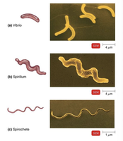
Spiral: Curved forms, including:
Vibrio: Comma-shaped, less than one turn
Spirillum: Helical, rigid, moves by flagella
Spirochete: Helical, flexible, moves by axial filament
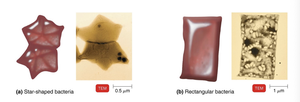
Less Common Shapes: Star-shaped and rectangular (often found in halophilic archaea)
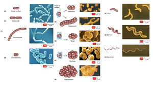
Arrangements
Pairs: Diplococci, diplobacilli
Clusters: Staphylococci
Chains: Streptococci, streptobacilli
Groups of Four: Tetrads
Cubelike Groups of Eight: Sarcinae
The Structure of a Prokaryotic Cell
Prokaryotic cells possess unique external and internal structures that contribute to their survival and pathogenicity.
Glycocalyx
Definition: A viscous, gelatinous sugar coat external to the cell wall, composed of polysaccharide and/or polypeptide.
Types:
Capsule: Well-organized and firmly attached to the cell wall.
Slime Layer: Loosely attached and unorganized.
Functions:
Contributes to virulence by preventing phagocytosis and aiding in adherence to surfaces.
Helps form biofilms (e.g., Streptococcus mutans in tooth decay).
Protects from dehydration.
Flagella
Structure: Composed of filament (flagellin), hook, and basal body.
Function: Provides motility, allowing bacteria to move toward or away from stimuli (taxis).
Arrangements:
Peritrichous: Flagella all around
Monotrichous: Single flagellum
Lophotrichous: Multiple flagella at one end
Amphitrichous: Flagella at both ends
Atrichous: No flagella
Movement:
Run: Counterclockwise rotation, straight movement
Tumble: Clockwise rotation, random change in direction
Stimuli: Chemotaxis (chemicals), phototaxis (light)
Axial Filaments
Also called endoflagella; found in spirochetes (e.g., Treponema pallidum, Borrelia burgdorferi).
Enable corkscrew movement.
Fimbriae and Pili
Fimbriae: Short, hairlike appendages for attachment; important in biofilm formation and pathogenicity (e.g., Neisseria gonorrhoeae).
Pili: Longer; involved in motility and DNA transfer (conjugation pili).
The Cell Wall
The cell wall is a semi-rigid structure that provides protection, maintains shape, and prevents osmotic lysis. Its composition is crucial for bacterial classification and pathogenicity.
Peptidoglycan: Main component, consisting of repeating units of N-acetylglucosamine (NAG) and N-acetylmuramic acid (NAM) linked by polypeptides.
Penicillin: Inhibits peptide cross-bridge formation, weakening the cell wall.
Gram-Positive Cell Walls
Thick peptidoglycan layer
Contains teichoic acids (lipoteichoic and wall teichoic acids)
Retains crystal violet dye (appears purple)
Provides antigenic specificity
Gram-Negative Cell Walls
Thin peptidoglycan layer
Three layers: outer membrane, cell wall, plasma membrane
Outer membrane contains lipopolysaccharide (LPS), lipoproteins, and phospholipids
LPS components: O polysaccharide (antigen), Lipid A (endotoxin)
Porins allow selective passage of molecules
More resistant to antibiotics and phagocytosis
Gram Stain Mechanism
Crystal violet-iodine complexes form inside cells
Alcohol dehydrates peptidoglycan in gram-positive cells (retains dye)
Alcohol dissolves outer membrane in gram-negative cells (dye washes out)
Safranin counterstain colors gram-negative cells pink/red
Atypical Cell Walls
Acid-fast cell walls: Thick peptidoglycan with mycolic acid (waxy lipid); stains red with carbolfuchsin (e.g., Mycobacterium, Nocardia).
Damage to the Cell Wall
Lysozyme: Hydrolyzes peptidoglycan bonds, weakening the wall.
Penicillin: Inhibits peptide bridge formation.
Protoplast: Wall-less gram-positive cell
Spheroplast: Wall-less gram-negative cell (retains outer membrane)
Both are susceptible to osmotic lysis.
The Plasma (Cytoplasmic) Membrane
The plasma membrane is a selectively permeable barrier composed of a phospholipid bilayer and proteins. It is essential for cellular metabolism and transport.
Fluid Mosaic Model: Describes the dynamic nature of the membrane, with proteins and phospholipids moving laterally.
Functions: Selective permeability, ATP production, enzymatic activity.
Movement of Materials Across Membranes
Passive Processes:
Simple diffusion: Movement from high to low concentration (e.g., O2, CO2).
Facilitated diffusion: Uses transport proteins for ions/large molecules.
Osmosis: Water movement across a selectively permeable membrane.
Osmotic pressure: Pressure needed to stop water movement.
Isotonic, hypotonic, hypertonic solutions affect water movement and cell integrity.
Active Processes:
Active transport: Requires ATP to move substances against the gradient.
Group translocation (prokaryotes only): Substance is chemically modified during transport (e.g., glucose to glucose-6-phosphate).
Cytoplasm
The cytoplasm is the internal matrix of the cell, containing water, proteins, carbohydrates, lipids, ions, and essential cellular structures.
Includes the nucleoid (DNA), ribosomes, and inclusions.
The Nucleoid
Bacterial chromosome: Circular, double-stranded DNA, not membrane-bound.
Plasmids: Small, circular DNA molecules; carry genes for antibiotic resistance and toxins; replicate independently.
Ribosomes
Sites of protein synthesis
Composed of protein and rRNA
70S ribosome: 50S (large) + 30S (small) subunits
Inclusions
Reserve deposits for nutrients
Types:
Metachromatic granules (phosphate reserves)
Polysaccharide granules (energy reserves)
Lipid inclusions (energy reserves)
Sulfur granules (energy reserves)
Carboxysomes (CO2 fixation)
Gas vacuoles (buoyancy)
Magnetosomes (iron oxide, destroy H2O2)
Endospores
Highly durable, dormant structures formed in response to adverse conditions
Resistant to desiccation, heat, chemicals, and radiation
Produced by Bacillus and Clostridium species
Sporulation: Formation of endospores
Germination: Return to vegetative state
Additional info: The above notes integrate foundational microbiology concepts with expanded academic context for clarity and exam preparation.
