 Back
Fundamentals of Microbial Growth and Decontamination Chapter 7 Notes: Study Guide
Study Guide - Smart Notes
 Tailored notes based on your materials, expanded with key definitions, examples, and context.
Fundamentals of Microbial Growth and Decontamination
Microbial Growth Basics
Microbial growth refers to the increase in cell number, not cell size, and is a fundamental process in microbiology. Growth can occur in laboratory conditions or in nature, with significant differences in complexity and environmental interactions.
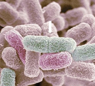
Binary Fission: Most prokaryotes divide by binary fission, a process where a single cell splits into two genetically identical daughter cells.
Budding and Spore Formation: Some microbes reproduce by budding (asexual outgrowth) or spore formation (asexual or sexual, depending on organism).
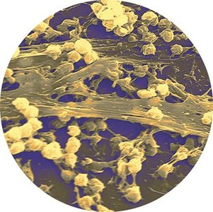
Biofilms: In nature, microbes often form biofilms—structured communities attached to surfaces, which are highly resistant to treatment and contribute to persistent infections.
Generation Time: The time required for a cell to divide. It varies by species and environmental conditions (e.g., E. coli: 20 minutes; Mycobacterium tuberculosis: 15–20 hours).
Bacterial Growth Curve
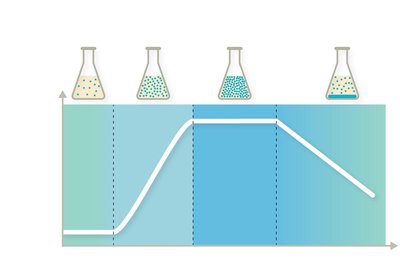
Bacteria grown in a closed batch system exhibit four distinct growth phases, which are important for both industrial and clinical microbiology.
Lag Phase: Cells adjust to their environment; no significant increase in cell number.
Log (Exponential) Phase: Rapid cell division and population growth.
Stationary Phase: Nutrient depletion and waste accumulation; cell division rate equals cell death rate.
Death Phase: Cells die as nutrients are exhausted and waste accumulates.
Industrial Microbial Growth Control
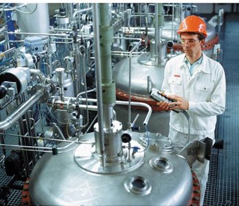
Maintaining cells at specific growth phases is crucial in industrial microbiology. Chemostats are used to continuously supply fresh nutrients and remove waste, keeping cultures in the desired phase.
Prokaryotic Growth Requirements
Temperature
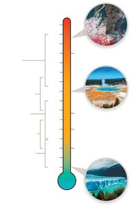
Temperature is a key environmental factor affecting microbial growth. Microbes are classified based on their optimal temperature ranges:
Psychrophiles: Grow at –20°C to 10°C.
Psychrotrophs: Grow at 0°C to 30°C; often associated with foodborne illness.
Mesophiles: Grow at 10°C to 50°C; most human pathogens fall in this group.
Thermophiles: Grow at 40°C to 75°C; found in compost piles and hot springs.
Extreme Thermophiles: Grow at 65°C to 120°C; often found in deep-sea vents.
pH
Microbes are also classified by their pH tolerance:
Acidophiles: Grow at pH 1–5; found in sulfur hot springs and volcanic vents.
Neutralophiles: Grow at pH 5–8; majority of microorganisms.
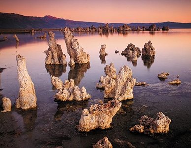
Alkaliphiles: Grow at pH 9–11; found in soda lakes.
Salinity
Halophiles thrive in high-salt environments (up to 35%), such as the Dead Sea and Great Salt Lake. Halotolerant microbes can survive higher salt concentrations but may not grow optimally.
Oxygen Requirements
Microbes are classified based on their oxygen use and tolerance:
Obligate Aerobes: Require oxygen for growth.
Obligate Anaerobes: Cannot tolerate oxygen.
Facultative Anaerobes: Can grow with or without oxygen.
Microaerophiles: Require low levels of oxygen.
Aerotolerant Anaerobes: Tolerate oxygen but do not use it.
Classification | Oxygen Use | Growth Pattern | ROS Management |
|---|---|---|---|
Obligate Aerobe | Absolute dependence | Top of tube | Yes |
Obligate Anaerobe | Not used | Bottom of tube | No |
Microaerophile | Small amounts | Middle of tube | Yes (low amounts) |
Aerotolerant Anaerobe | Not used | Throughout tube | Yes |
Facultative Anaerobe | Prefer oxygen, can survive without | Throughout tube, dense at top | Yes |
Microbial Nutrition and Energy Sources
Essential Nutrients and Growth Factors
Microbes require essential nutrients to build cellular components. These include macronutrients (needed in large amounts) and micronutrients (needed in small amounts).
Macronutrients: Carbon, hydrogen, oxygen, nitrogen.
Micronutrients: Iron, copper, zinc, etc.
Growth Factors: Substances microbes cannot synthesize (e.g., amino acids, vitamins).
Energy Sources
Phototrophs: Use light energy.
Chemotrophs: Use energy from chemical compounds.
Autotrophs: Use inorganic carbon (CO2).
Heterotrophs: Require organic carbon.
Growing, Isolating, and Counting Microbes
Culture Media
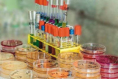
Microbes are grown on various media, classified by physical state, chemical composition, and function.
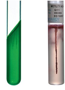
Physical State: Liquid (broth), solid (agar plates), semisolid (motility testing).
Chemical Composition: Defined (precisely known), complex (unknown exact composition).
Function: Selective (favors certain microbes), differential (distinguishes microbes visually).
Differential and Selective Media
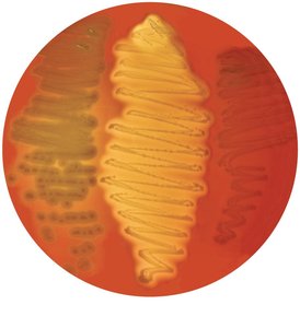
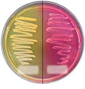
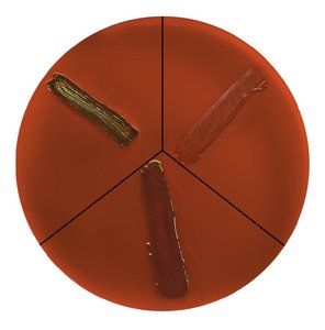
Blood agar is a differential medium that distinguishes microbes based on hemolysis. Mannitol salt agar and eosin methylene blue agar are selective and differential media used to isolate and differentiate bacteria.
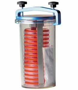
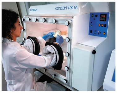
Anaerobic Culture Methods
Anaerobic media and chambers are used to culture microbes that cannot tolerate oxygen.
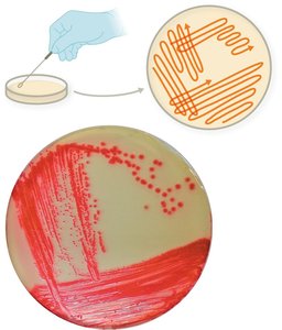
Streak Plate Technique
The streak plate technique is used to isolate pure cultures by spreading cells thinly across an agar surface.
Methods for Counting Microbes
Microbial enumeration is essential in clinical and industrial settings. Methods are classified as direct or indirect.
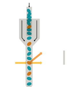
Direct Methods: Manual cell counts, automated cell counts (Coulter counter, flow cytometer), viable plate counts.
Indirect Methods: Turbidity measurement (spectrophotometry), dry weight, biochemical activity.
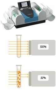
Method Name | Description |
|---|---|
Microscopic count | Manual count of cells using a microscope and counting chamber; does not differentiate living/dead cells |
Coulter counter/flow cytometer | Automated methods; flow cytometer distinguishes cells by fluorescent tags |
Viable plate count | Colonies from diluted sample reflect original cell count |
Turbidity measurement | Spectrophotometer measures cloudiness of culture |
Dry weight | Dry mass measurement |
Biochemical activity | Byproducts of cell growth measured |
Controlling Microbial Growth
Physical Methods
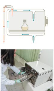
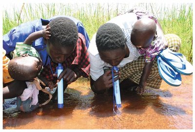
Physical methods include temperature changes, radiation, and filtration.
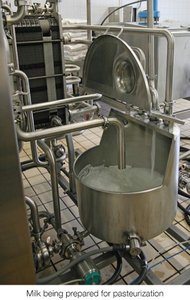
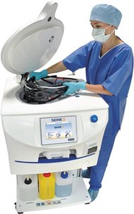
Heat: Sterilization (autoclaving, dry heat), decontamination (boiling, pasteurization).
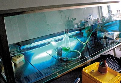
Radiation: Ionizing (gamma rays, X-rays) and nonionizing (UV light).
Filtration: HEPA filters for air, membrane filters for liquids.
Chemical Methods (Germicides)
Chemical control involves germicides, which are classified by their effectiveness and application.
Disinfectants: Used on inanimate objects.
Antiseptics: Used on living tissue.
Germicide Levels: Low (detergents), intermediate (alcohols, phenols), high (aldehydes, halogens, peroxygens, ethylene oxide).
Level | Germicide | Mode of Action | Pros/Cons |
|---|---|---|---|
Low | Detergents | Target lipid membranes | Cheap, low toxicity; activity decreased in hard water |
Intermediate | Alcohols | Target proteins and lipid membranes | Cheap, flammable |
Intermediate | Phenols | Target proteins and lipid membranes | Effective in hard water, irritant |
High | Aldehydes | Target proteins, nucleic acids | Toxic, achieve sterility |
High | Halogens | Oxidizing agents | Cheap, sterilant at high concentration |
High | Peroxygens | Oxidizing agents | Effective sterilization, corrosive |
High | Ethylene oxide | Targets proteins, nucleic acids | Gentle on equipment, toxic |
Microbial Control for Specific Microbes
Mycobacteria: Controlled by reducing airborne particles.
Endospores: Eliminated by autoclaving or sporicides.
Viruses: Envelope viruses are sensitive to heat and detergents; naked viruses to chlorine.
Protozoa: Controlled by filtration, UV, ozone.
Prions: Require combined chemical and physical treatments.
Clinical Case Example: Infection Control
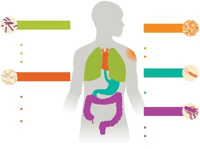
Case studies highlight the importance of proper decontamination and aseptic techniques in preventing infections, such as those caused by Pseudomonas aeruginosa in ear piercings.
Visual Summary
Additional info: This study guide expands on brief points from the original material, providing definitions, examples, and context for each topic. Tables are recreated for clarity and completeness. Images are included only when directly relevant to the adjacent content.
