 Back
BackFungi: Structure, Nutrition, Reproduction, and Classification
Study Guide - Smart Notes
 Tailored notes based on your materials, expanded with key definitions, examples, and context.
Fungi: An Overview
What Are Fungi?
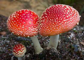
Fungi are a distinct kingdom of eukaryotic organisms, separate from plants, animals, and bacteria. They include yeasts, molds, and mushrooms, and are widely distributed across terrestrial and aquatic ecosystems. Fungi play essential roles in decomposition, symbiosis, and disease.
Kingdom Fungi: Eukaryotic, non-photosynthetic organisms.
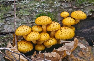
Major Groups: Yeasts (unicellular), molds (filamentous), mushrooms (fruiting bodies).
Distribution: Found in soil, water, air, and as symbionts or pathogens.
Example: Mushrooms are a common macroscopic form of fungi.
Structural Characteristics of Fungi
Cell Wall and Growth Forms
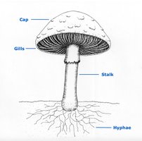
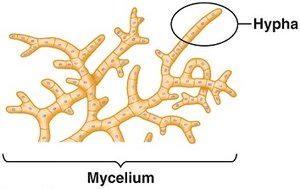
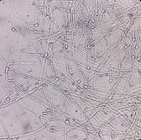
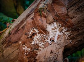
Fungal cells possess cell walls containing chitin, a polymer that provides rigidity and protection. Most fungi grow as hyphae—long, branching filamentous cells that collectively form a mycelium. Some fungi, such as yeasts, exist as unicellular forms.
Chitin: Main structural component of fungal cell walls (not cellulose).
Hyphae: Thread-like filaments; basic structural unit.
Mycelium: Interconnected network of hyphae; increases surface area for nutrient absorption.
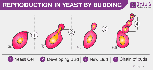
Yeasts: Unicellular fungi; reproduce by budding.
Fungal Nutrition
Absorptive Nutrition
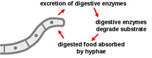
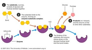
Fungi utilize absorptive nutrition, secreting enzymes to digest food externally and then absorbing the resulting small, soluble molecules. As heterotrophs, fungi must obtain organic molecules from their environment for energy and carbon.
Exoenzymes: Secreted enzymes (cellulases, proteases, lipases) break down complex molecules.
External Digestion: Digestion occurs outside the fungal body.
Absorption: Nutrients are absorbed through hyphal cell walls and membranes.
Modes of Fungal Nutrition
Saprotrophs: Obtain nutrients from dead and decaying organic matter; act as decomposers.
Parasites: Live in or on living hosts; use haustoria to penetrate host cells and absorb nutrients.
Mutualists: Form beneficial symbiotic relationships (e.g., mycorrhizae with plant roots).
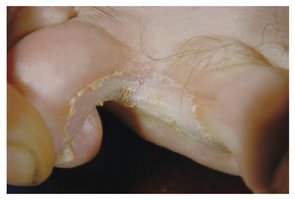
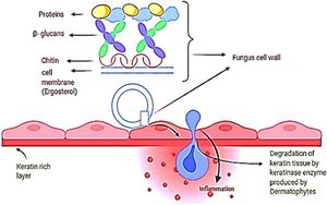
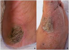
Fungal Pathogenesis: Athlete’s Foot
Athlete’s foot is a contagious fungal infection caused by dermatophytes that digest keratin in the skin, leading to irritation and breakdown. Dermatophytes secrete keratin-degrading enzymes, and prefer dead, keratinized skin layers.
Keratin: Structural protein in skin, hair, and nails.
Enzymes: Proteases (e.g., fungalysin) break down keratin.
Spread: May infect toenails (onychomycosis) if untreated.


Fungal Hyphae
Structure and Function
Fungal hyphae are microscopic, thread-like filaments composed of chitin. They grow by apical extension, enabling efficient nutrient absorption. Hyphae form the mycelium, which is the vegetative body of filamentous fungi.
Nutrient Absorption: Release enzymes and absorb nutrients.
Structural Support: Form mycelium for vegetative growth.
Reproduction: Specialized hyphae produce and disperse spores.
Symbiosis: Mycorrhizal hyphae connect with plant roots.
Pathogenesis: Haustoria penetrate host tissues.
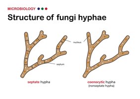
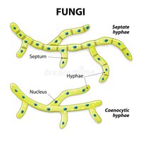
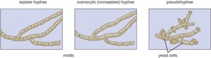
Types of Hyphae
Septate Hyphae: Divided by septa (cross-walls) with pores for cytoplasmic flow.
Coenocytic (Aseptate) Hyphae: Lack septa; contain multiple nuclei in a shared cytoplasm.
Pseudohyphae: Chains of elongated yeast cells attached after division; constrictions at junctions.

Hyphae Structure and Composition
Chitin: Provides rigidity and protection.
Apical Growth: Growth occurs at hyphal tips.
Diameter: Typically 2–10 µm.

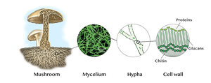
Fungal Cell Wall Structure
Inner Layer (Structural)
The inner layer of the fungal cell wall provides mechanical strength and rigidity. It is composed primarily of chitin and β-glucans, which are cross-linked to form a pressure-resistant mesh.
Chitin: Long chains of N-acetylglucosamine.
β-glucans: Glucose polymers.

Outer Layer (Interactive)
The outer layer is enriched in mannoproteins, which facilitate cell–cell recognition, adhesion, and immune evasion.
Mannoproteins: Proteins with mannan sugar chains.
Functions: Adhesion, immune evasion, environmental interactions.
Fungal Reproduction
Asexual Reproduction
Asexual reproduction is the most common mode, enabling rapid colonization under favorable conditions. Offspring are genetically identical to the parent.
Sporogenesis: Formation of mitospores (conidia) by hyphae.
Budding: Common in yeasts; new cell forms and separates from parent.
Fragmentation: Mycelium breaks and fragments grow into new colonies.
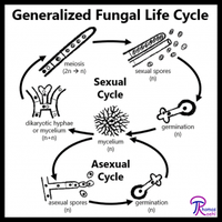
Sexual Reproduction
Sexual reproduction is initiated under harsh conditions, generating genetic variation for adaptability. Most fungi have mating types rather than distinct sexes.
Plasmogamy: Fusion of cytoplasm from two parent cells.
Karyogamy: Fusion of haploid nuclei to form diploid zygote.
Meiosis: Diploid nucleus divides to restore haploid state, producing diverse spores.
Classification of Fungi
Major Phyla
Fungi are classified based on sexual reproduction, molecular phylogeny, and hyphal structure. The main phyla include:
Phylum | Key Features | Example |
|---|---|---|
Chytridiomycota | Aquatic, motile zoospores with flagella | Chytrids |
Zygomycota (historical) | Coenocytic hyphae, zygospore formation | Bread molds |
Glomeromycota | Arbuscular mycorrhizal symbionts | Mycorrhizal fungi |
Ascomycota | Sexual spores in asci; yeasts, molds, pathogens | Saccharomyces, Aspergillus |
Basidiomycota | Basidiospores on basidia; mushrooms, rusts, smuts | Agaricus, Puccinia |
Deuteromycota (obsolete) | No observed sexual stage; reclassified as sexual cycles discovered | Penicillium |
Review Questions
Which feature best distinguishes fungi from plants? C. Use of absorptive nutrition
Why is fungal digestion considered “external”? B. Enzymes act outside the fungal body
Haustoria are specialized fungal structures primarily involved in: C. Host penetration and nutrient absorption
The primary structural polymer in fungal cell walls is: C. Chitin
Which hyphal type lacks septa and contains multiple nuclei within a shared cytoplasm? C. Coenocytic hyphae
The outer layer of the fungal cell wall is primarily involved in: C. Environmental and host interactions
Which reproductive strategy produces genetically identical offspring? C. Asexual reproduction
Which asexual mechanism is most common in yeasts? C. Budding
Why do fungi often switch to sexual reproduction under harsh conditions? C. It increases genetic variation
