 Back
BackGenetic Variation and Microbial Evolution: Mechanisms and Consequences
Study Guide - Smart Notes
 Tailored notes based on your materials, expanded with key definitions, examples, and context.
Genetic Variation and Microbial Evolution
Introduction to Microbial Genome Evolution
Microbial genomes are highly dynamic and constantly evolving, which is essential for the diversification of the microbial world. Evolution in microbes can occur through the loss or acquisition of genes, such as the evolution of pathogenic Shigella from non-pathogenic E. coli by gene loss, or the acquisition of virulence genes by enteropathogenic E. coli from the environment.
Genetic Variation in Eukaryotes
Mechanisms of Genetic Variation
Genetic variation in eukaryotes is primarily generated through three mechanisms during sexual reproduction:
Independent assortment of homologous chromosomes: Chromosomes are randomly distributed into gametes, resulting in genetic diversity among offspring.
Crossing over: Homologous chromosomes exchange genetic material during meiosis, producing recombinant chromosomes.
Random fertilization: The combination of gametes from two parents further increases genetic diversity.
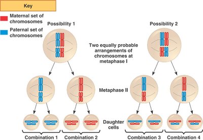
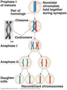
Vertical gene transfer refers to the transfer of genes from parents to progeny, explaining why siblings do not look identical.
Genetic Variation in Prokaryotes
Mechanisms of Genetic Variation
Prokaryotes do not undergo sexual reproduction like eukaryotes, but they generate genetic diversity through:
Mutations: Stable, heritable changes in the genome sequence.
Horizontal Gene Transfer (HGT): Transfer of genetic material between unrelated organisms, which is crucial for microbial evolution.
HGT can occur via three main mechanisms:
Transformation: Uptake of naked DNA from the environment by competent cells.
Transduction: Transfer of bacterial genes by bacteriophages (bacterial viruses).
Conjugation: Direct transfer of DNA from one bacterium to another via cell-to-cell contact, often mediated by plasmids.
Transformation
Transformation involves the uptake and incorporation of naked DNA from the environment into a recipient cell, resulting in heritable genetic changes. Only competent cells can undergo transformation.
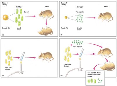
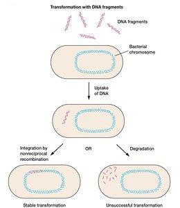
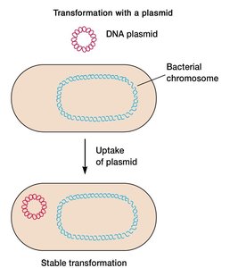
Some bacteria are naturally competent (e.g., Streptococcus pneumoniae), while others can be made competent through chemical or electrical treatments.
Plasmids
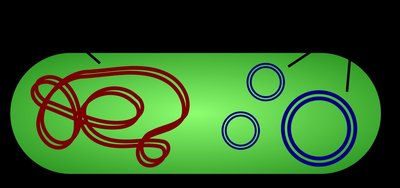
Plasmids are small, double-stranded, circular DNA molecules that can exist independently of the bacterial chromosome. They often carry genes that confer selective advantages, such as antibiotic resistance (R plasmids), virulence, or metabolic functions.
Transduction
Transduction is the process by which bacterial DNA is transferred from one bacterium to another by a bacteriophage. A transducing particle is a phage that carries host cell DNA instead of its own viral DNA.
Conjugation
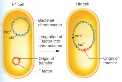
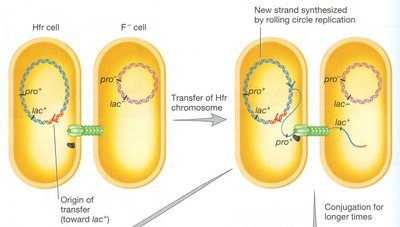
Conjugation is the transfer of DNA from a donor to a recipient bacterium via direct cell-to-cell contact, typically mediated by a pilus and conjugative plasmids such as the F (fertility) factor in E. coli.
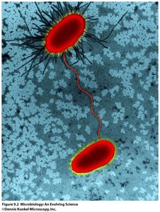
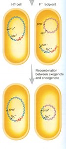
Conjugative plasmids carry genes for pilus formation and DNA transfer. The F factor can integrate into the bacterial chromosome, creating an Hfr (high frequency of recombination) strain, which can transfer chromosomal genes during conjugation.
Experimental Evidence for Conjugation
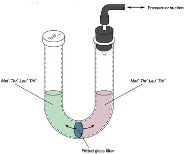
Tatum and Lederberg demonstrated conjugation using double auxotrophs and minimal media to select for prototrophic recombinants. Bernard Davis's U-tube experiment showed that direct cell-to-cell contact is necessary for conjugation.

Mutations and Their Effects
Types and Causes of Mutations
Mutations are stable, heritable changes in the genome sequence. They can be:
Neutral: No effect on phenotype.
Detrimental: Harmful to the organism.
Beneficial: Advantageous and likely to be selected for.
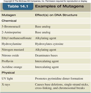
Mutations can arise spontaneously (e.g., replication errors, DNA damage, transposon insertion) or be induced by mutagens (physical or chemical agents).
Types of Mutations
Point mutations: Single base changes, which may be silent, missense, or nonsense.
Frameshift mutations: Insertions or deletions that alter the reading frame.
Strand breaks and crosslinks: Can lead to chromosomal rearrangements or loss of genetic information.
Mutant Phenotypes
Loss of function: Decreased or eliminated activity (e.g., a protein that can no longer bind its ligand).
Gain of function: Increased or new activity (e.g., a protein that is active without its normal regulator).
Reversions and Suppressor Mutations
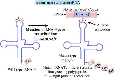
Reverse or back mutations restore the wild-type phenotype. Suppressor mutations can occur within the same gene (intragenic) or in a different gene (extragenic), compensating for the original mutation.
Mutations in Regulatory Sequences
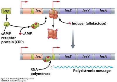
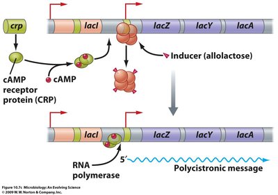
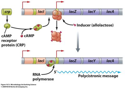
Mutations in non-coding regulatory regions can affect gene expression, such as mutations in the operator or promoter regions of the lac operon, impacting the binding of repressors or activators like CAP.
Screening for Mutants
Detection Methods
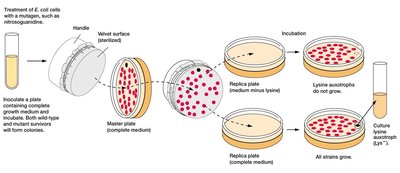
Because mutations are rare, sensitive detection methods are required. Replica plating is a common technique for identifying auxotrophs (e.g., lysine auxotrophs).
The Ames Test for Carcinogenicity
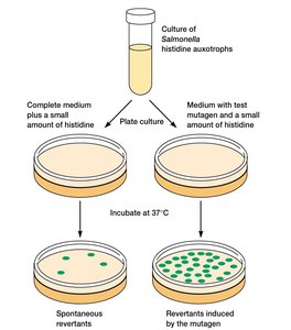
The Ames test screens for mutagenicity (and thus potential carcinogenicity) by measuring the reversion rate of histidine auxotrophs of Salmonella in the presence of a suspected mutagen. An increased reversion rate indicates mutagenic and possibly carcinogenic potential.


Mutagen | Effect(s) on DNA Structure |
|---|---|
5-Bromouracil | Base analog |
2-Aminopurine | Base analog |
Ethyl methanesulfonate | Alkylating agent |
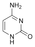
Hydroxylamine | Hydroxylates cytosine |
Nitrogen mustard | Alkylating agent |
Nitrous oxide | Deaminates bases |
Proflavin | Intercalating agent |
Acridine orange | Intercalating agent |
UV light | Promotes pyrimidine dimer formation |
X rays | Causes base deletions, single-strand nicks, cross-linking, and chromosomal breaks |
Additional info: The Ames test uses a "reverse mutation" screen because it is easier to detect the restoration of function (e.g., growth without histidine) than the loss of function, and most carcinogens are mutagens.
