 Back
Gram Stain: Principles, Procedure, and Interpretation
Study Guide - Smart Notes
 Tailored notes based on your materials, expanded with key definitions, examples, and context.
Gram Stain: Principles, Procedure, and Interpretation
Background and Significance
The Gram stain is a fundamental differential staining technique in microbiology, developed by Hans Christian Gram in 1884. It is used to distinguish between Gram-positive and Gram-negative bacteria based on differences in their cell wall structure. This method is clinically significant for guiding initial diagnosis and treatment of bacterial infections.
Differential stains allow for classification of bacteria beyond simple morphology, based on cell wall chemistry.
Gram-positive bacteria retain the primary stain and appear purple.
Gram-negative bacteria lose the primary stain and appear pink or red after counterstaining.
Principles of Gram Stain
The Gram stain differentiates bacteria by the thickness of their peptidoglycan layer and the presence or absence of an outer membrane.
Gram-positive bacteria: Thick peptidoglycan layer, no outer membrane.
Gram-negative bacteria: Thin peptidoglycan layer, outer membrane present.
The chemistry of the cell wall determines the retention or loss of the crystal violet-iodine complex during the decolorization step.
Steps of the Gram Stain Procedure
The Gram stain consists of four main steps, each with a specific function:
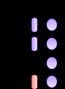
1. Primary stain (Crystal violet): Stains all cells purple initially.
2. Mordant (Gram’s iodine): Forms a complex with crystal violet (CV-I), enhancing retention in cells.
3. Decolorization (Alcohol): Alcohol dissolves the outer membrane of Gram-negative bacteria, allowing the CV-I complex to be washed out. In Gram-positive bacteria, alcohol dehydrates the thick peptidoglycan, trapping the CV-I complex.
4. Counterstain (Safranin): Stains decolorized Gram-negative cells pink/red; Gram-positive cells remain purple.
Example: Staphylococcus epidermidis (Gram-positive) will appear purple, while Escherichia coli (Gram-negative) will appear pink/red after staining.
Materials Required
Gram Stain Kit: Crystal violet, Gram’s iodine, ethanol, safranin, spray water bottle
Bacterial cultures: Staphylococcus epidermidis, Escherichia coli
Staining tray
Detailed Gram Stain Procedure
Follow these steps to perform a Gram stain:
Prepare bacterial smears on a slide (adjacent or mixed for best results).
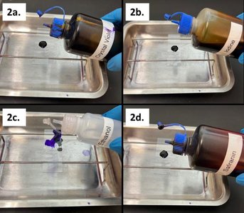
Stain the heat-fixed smears:
Cover with crystal violet for 1 minute, rinse with water.
Cover with Gram’s iodine for 1 minute, rinse with water.
Decolorize with ethanol for 10 seconds or until runoff is clear, rinse with water.
Cover with safranin for 45 seconds, rinse with water.
Blot dry using bibulous paper.
Observe under oil immersion microscopy.
Interpretation of Gram Stain Results
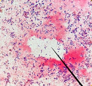
After staining, observe the slide under a microscope:
Gram-positive cocci: Purple
Gram-negative rods: Pink/red
Cell morphology (shape), arrangement, and Gram reaction are recorded for each specimen.
Clinical Significance of Gram Stain
The Gram stain is essential in clinical microbiology for:
Rapid identification of bacterial pathogens
Guiding initial antibiotic therapy
Distinguishing between different types of infections
Comparison: Gram-Positive vs. Gram-Negative Bacteria
The following table summarizes key differences:
Feature | Gram-Positive | Gram-Negative |
|---|---|---|
Peptidoglycan Layer | Thick | Thin |
Outer Membrane | Absent | Present |
Stain Color | Purple | Pink/Red |
Teichoic Acids | Present | Absent |
Lipopolysaccharide | Absent | Present |
Common Questions and Troubleshooting
If the counterstain (Safranin) is omitted:
Gram-positive bacteria remain purple.
Gram-negative bacteria appear colorless or faintly stained.
Mixed Gram reactions in a pure culture: May indicate culture age, improper technique, or contamination.
Gram stain is a differential stain: True.
Gram stain is best performed on fresh cultures: False; older cultures may yield unreliable results.
Summary Table: Gram Stain Steps
Step | Reagent | Purpose |
|---|---|---|
1 | Crystal violet | Primary stain (colors all cells purple) |
2 | Gram’s iodine | Mordant (forms CV-I complex) |
3 | Ethanol | Decolorizer (removes CV-I from Gram-negative cells) |
4 | Safranin | Counterstain (colors Gram-negative cells pink/red) |
Key Terms
Peptidoglycan: Structural polymer in bacterial cell walls.
Mordant: Substance that fixes a dye in cells.
Decolorizer: Removes stain from certain cells based on cell wall properties.
Counterstain: Secondary stain to color decolorized cells.
Equations and Scientific Principles
While the Gram stain is not based on mathematical equations, the process relies on chemical interactions:
Formation of the crystal violet-iodine complex:
Retention or loss of CV-I complex depends on cell wall structure and alcohol treatment.
Additional info: The Gram stain is a foundational technique for bacterial classification and is often the first step in clinical microbiology labs for identifying pathogens.
