 Back
BackHistory of Microbiology and Microscopy: Foundations, Tools, and Key Concepts
Study Guide - Smart Notes
 Tailored notes based on your materials, expanded with key definitions, examples, and context.
History of Microbiology
Introduction to Microbes
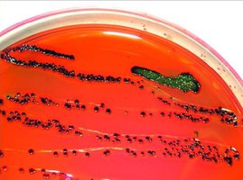
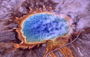
Microbiology is the study of organisms too small to be seen without magnification. These include bacteria, archaea, fungi, protozoa, helminths, viruses, and prions. Microbes are found everywhere there is sufficient moisture, and some thrive in extreme environments.
Bacteria and Archaea: Unicellular, lack nuclei, reproduce asexually, and are found in diverse environments, including extreme conditions.
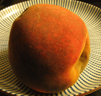
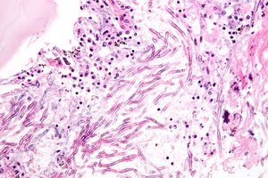
Fungi: Eukaryotic, possess cell walls, obtain food from other organisms. Includes molds (multicellular, hyphae, spores) and yeasts (unicellular, budding).
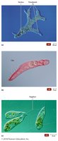
Protozoa: Single-celled eukaryotes, similar to animals, capable of locomotion, reproduce asexually or sexually.
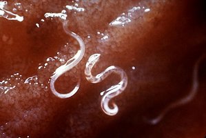
Helminths: Parasitic worms, eukaryotic, multicellular, with microscopic egg and larval stages.
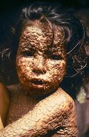
Viruses: Acellular, obligate intracellular parasites, much smaller than bacteria, cause a wide range of diseases.
Prions: Protein-only infectious agents, cause diseases such as mad cow disease and Creutzfeldt-Jakob Disease.
Development of Microbiology: Key Historical Experiments
The field of microbiology was shaped by experiments that challenged ancient beliefs and established scientific principles.
Spontaneous Generation (Abiogenesis): The ancient belief that life arises from non-living matter, disputed by biogenesis.
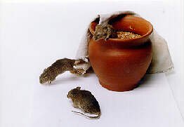
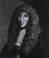
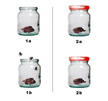
Redi’s Experiments: Demonstrated that maggots do not spontaneously arise from meat, disproving abiogenesis.
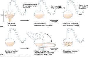
Pasteur’s Experiments: Used swan-neck flasks to show that microbes do not spontaneously generate, supporting biogenesis and leading to the germ theory of disease.
Major Contributors to Microbiology
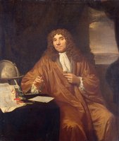
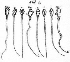
Antonie van Leeuwenhoek: Developed powerful microscopes, discovered protozoa and bacteria, considered the father of bacteriology and protozoology.
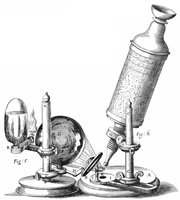
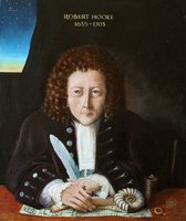
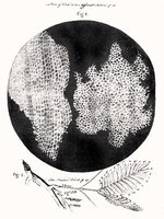
Robert Hooke: First described cells in cork, published the first description of a microorganism (fungus), contributed to cell theory.
Louis Pasteur: Disproved spontaneous generation, developed pasteurization, initiated germ theory, created vaccines for rabies and anthrax.
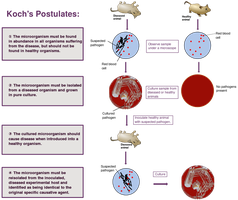
Robert Koch: Proved germ theory, isolated causative agents of diseases, developed Koch’s postulates for identifying pathogens.
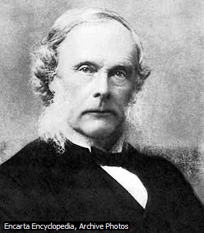
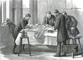
Joseph Lister: Introduced aseptic techniques in surgery, reducing infection rates.
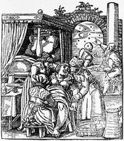
Ignaz Semmelweis: Advocated handwashing to prevent puerperal fever.
Microscopy and Microbiological Tools
Microscopy: Principles and Types
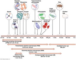
Microscopy is essential for studying microorganisms. It relies on magnification, resolution, contrast, and the use of stains to visualize cells and their structures.
Magnification: Enlargement of an image.
Resolution: Ability to distinguish two points as separate.
Contrast: Difference in light intensity between specimen and background.
Wavelength: Shorter wavelengths yield higher resolution.
Types of Microscopes
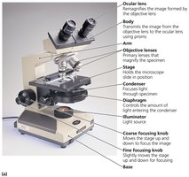
Light Microscopes: Includes bright-field, simple, and compound microscopes.
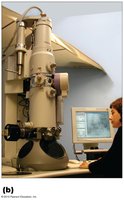
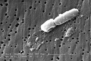
Electron Microscopes: Transmission Electron Microscope (TEM) and Scanning Electron Microscope (SEM) offer much higher magnification and resolution, allowing visualization of cell ultrastructure.
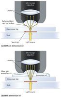
Immersion Oil and Resolution
Immersion oil increases resolution by reducing light refraction between the specimen and the objective lens.
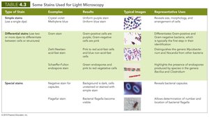
Staining Techniques
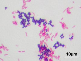
Staining enhances contrast and reveals cellular features. Stains are classified as basic (cationic) or acidic (anionic) dyes. Fixed, stained smears are prepared by spreading a thin film of cells, air-drying, and heat-fixing.
Simple Stains: Use a single dye to highlight cell shape and arrangement.
Differential Stains: Use multiple dyes to distinguish cell types or structures (e.g., Gram stain, acid-fast stain, endospore stain).
Special Stains: Reveal specific structures (e.g., capsule, flagella).
Bacterial Morphology and Arrangement
Bacteria exhibit a variety of shapes and arrangements, which are important for identification and classification.
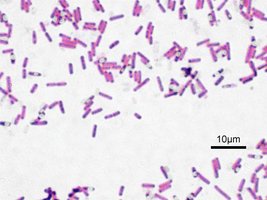
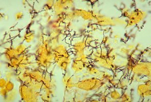
Shapes: Coccus (spherical), Bacillus (rod-shaped), Coccobacillus (short and plump), Vibrio (curved), Spirillum (spiral), Spirochete (corkscrew), Pleomorphism (variable shapes).
Arrangements: Cocci (single, pairs, tetrads, clusters, chains, cubical packets), Bacilli (single, pairs, chains, palisades).
Key Concepts and Take Home Points
Understand the contributions of major scientists to microbiology.
Be able to design experiments proving biogenesis.
Know the principles of germ theory, Koch’s postulates, antiseptic technique, and fermentation.
Identify types of microscopes and their limits.
Recognize bacterial morphology and arrangements.
Differentiate staining methods and their applications.
Additional info:
Pasteurization is a process developed by Louis Pasteur to reduce microbial load in food and beverages by heating.
Koch’s postulates are foundational for linking specific microbes to specific diseases.
Electron microscopy allows visualization of viruses and cell organelles, which are beyond the resolution of light microscopes.
