 Back
BackHost Defenses and Innate Immunity: An Overview of the Immune System
Study Guide - Smart Notes
 Tailored notes based on your materials, expanded with key definitions, examples, and context.
Overview of Host Defense Mechanisms
Introduction to Host Defenses
The human body is equipped with a complex network of defense mechanisms to protect against pathogens. These mechanisms are categorized into innate (nonspecific) and adaptive (specific) immunity. Innate defenses are present at birth and provide immediate, nonspecific protection, while adaptive immunity develops in response to specific pathogens and provides long-lasting protection through immunological memory.


Lines of Defense
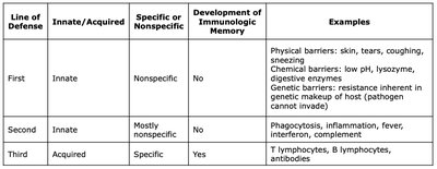
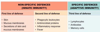
The immune system operates through three main lines of defense, each with distinct characteristics and roles in host protection.
Line of Defense | Innate/Acquired | Specific or Nonspecific | Development of Immunologic Memory | Examples |
|---|---|---|---|---|
First | Innate | Nonspecific | No | Physical barriers: skin, tears, coughing, sneezing Chemical barriers: low pH, lysozyme, digestive enzymes Genetic barriers: resistance inherent in genetic makeup of host |
Second | Innate | Mostly nonspecific | No | Phagocytosis, inflammation, fever, interferon, complement |
Third | Acquired | Specific | Yes | T lymphocytes, B lymphocytes, antibodies |
First Line of Defense: Innate, Nonspecific Barriers
Physical or Anatomical Barriers
The first line of defense consists of physical and anatomical barriers that prevent the entry of pathogens. These barriers are always present and act nonspecifically against a wide range of invaders.
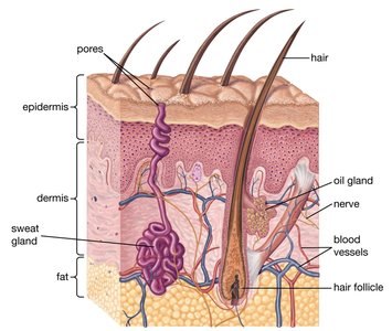
Skin: The outermost layer of skin is composed of tightly packed epithelial cells filled with keratin, making it a formidable barrier. Sweat and sebaceous secretions further inhibit microbial growth.
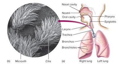
Mucous Membranes: Line the digestive, respiratory, and genitourinary tracts. Mucus traps microbes, and ciliary action in the respiratory tract helps expel them.
Mechanical Actions: Blinking, tear production, saliva flow, urination, defecation, and vomiting help flush out pathogens.
Chemical Barriers
Chemical defenses are produced by the skin and mucous membranes to inhibit or destroy pathogens.
Sebum: Lubricates the skin and creates an inhospitable environment for microbes.
Lysozyme: An enzyme found in tears and saliva that hydrolyzes bacterial cell walls.
Defensins: Antimicrobial peptides that disrupt microbial membranes.
Acidic pH: Sweat, stomach acid, and the vaginal tract maintain low pH environments that inhibit microbial growth.
Genetic Barriers
Genetic factors can determine susceptibility or resistance to certain infections. Some species or individuals possess genetic traits that prevent infection by specific pathogens.
Species Specificity: Many pathogens infect only certain species due to receptor compatibility.
Genetic Polymorphisms: For example, individuals with sickle-cell trait are resistant to malaria.

Second Line of Defense: Innate, Mostly Nonspecific Responses
Cellular and Chemical Defenses
If pathogens breach the first line of defense, the second line provides immediate, nonspecific responses. This includes the action of various white blood cells (WBCs), inflammation, fever, and antimicrobial proteins.
Phagocytic Leukocytes: Neutrophils, eosinophils, and macrophages ingest and destroy pathogens.
Inflammatory Response: Localized response to injury or infection, characterized by redness, heat, swelling, and pain.
Fever: Systemic increase in body temperature that inhibits pathogen growth and enhances immune activity.
Antimicrobial Proteins: Interferons and complement proteins disrupt pathogen replication and facilitate immune cell recruitment.
Major Components of the Immune System
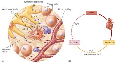
The immune system is composed of several interconnected compartments, including the reticuloendothelial system (RES), extracellular fluid (ECF), blood, and lymphatic system. Effective immune responses require communication between these compartments.
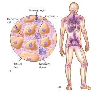
RES: Network of connective tissue fibers and phagocytic cells (mainly macrophages) that filter pathogens from tissues.
Blood: Contains plasma, serum, erythrocytes, leukocytes, and platelets.
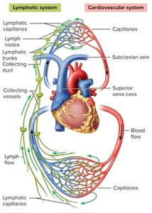
Lymphatic System: Returns extracellular fluid to the bloodstream and provides sites for immune surveillance and response.
Blood and Hematopoiesis
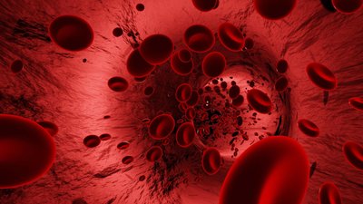
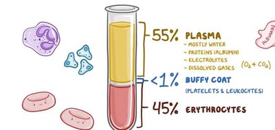
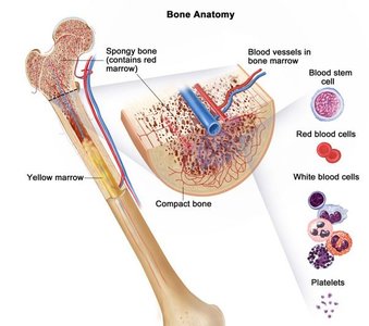
Blood is a vital fluid composed of plasma, erythrocytes (RBCs), leukocytes (WBCs), and platelets. Hematopoiesis is the process by which all blood cells are produced from stem cells in the bone marrow.
Plasma: The liquid portion of blood, containing water, proteins, electrolytes, and other solutes.
Serum: Plasma without clotting factors.
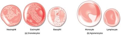
Leukocytes: Divided into granulocytes (neutrophils, eosinophils, basophils) and agranulocytes (lymphocytes, monocytes).
Leukocytes: Granulocytes and Agranulocytes
Leukocytes are the primary cells involved in immune responses. They are classified based on the presence or absence of cytoplasmic granules.
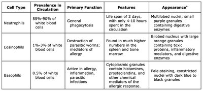
Cell Type | Prevalence in Circulation | Primary Function | Features | Appearance |
|---|---|---|---|---|
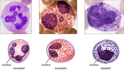
Neutrophils | 55%-90% | General phagocytosis | Short lifespan, rapid responders | Multilobed nuclei, small granules |
Eosinophils | 1%-3% | Destruction of parasitic worms, allergy mediators | Bilobed nucleus, orange granules | Large granules, digestive enzymes |
Basophils | 0.5% | Allergy, inflammation, parasitic infections | Histamine-containing granules | Constricted nucleus, dark granules |
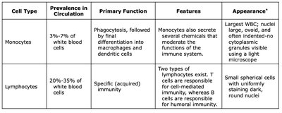
Cell Type | Prevalence in Circulation | Primary Function | Features | Appearance |
|---|---|---|---|---|
Monocytes | 3%-7% | Phagocytosis, differentiation into macrophages and dendritic cells | Secrete immune-modulating chemicals | Largest WBC, oval nucleus |
Lymphocytes | 20%-35% | Specific (acquired) immunity | B cells (antibody production), T cells (cell-mediated immunity) | Small, round nucleus |
Origin and Function of Blood Cells
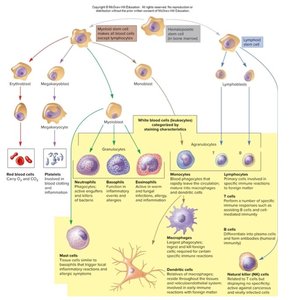
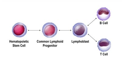
All blood cells originate from hematopoietic stem cells in the bone marrow. These stem cells differentiate into various lineages, giving rise to erythrocytes, leukocytes, and platelets.
Lymphatic System
Structure and Function
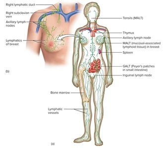
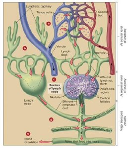
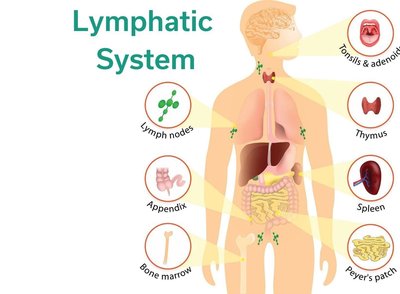
The lymphatic system is a network of vessels, organs, and tissues that helps maintain fluid balance, filters pathogens, and supports immune responses. Lymphatic fluid (lymph) is derived from blood plasma and circulates through lymphatic vessels, eventually returning to the bloodstream.
Lymph Nodes: Filter lymph and provide sites for immune cell activation.
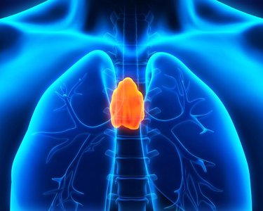
Thymus: Site of T cell maturation.
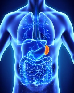
Spleen: Filters blood, removes old RBCs, and detects pathogens.
MALT, GALT, SALT: Collections of lymphoid tissue in mucosal, gut, and skin regions.
Second Line of Defense: Mechanisms
Inflammatory Response
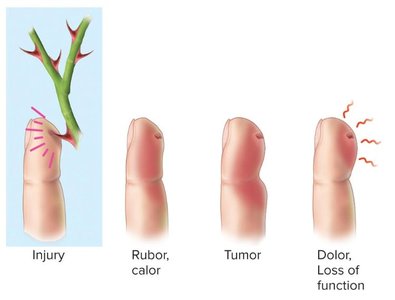
Inflammation is a localized, nonspecific response to tissue injury or infection. It serves to contain and eliminate pathogens, remove damaged cells, and initiate tissue repair.
Redness (Rubor): Increased blood flow to the area.
Heat (Calor): Elevated temperature due to increased circulation.
Swelling (Tumor): Accumulation of fluid (edema).
Pain (Dolor): Stimulation of nerve endings.
Phagocytosis
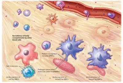
Phagocytosis is the process by which certain cells (phagocytes) engulf and digest pathogens and debris. Major phagocytes include neutrophils, eosinophils, and macrophages. Dendritic cells also play a role in presenting antigens to lymphocytes.
Recognition: Phagocytes recognize pathogens via pattern recognition receptors (PRRs) that bind pathogen-associated molecular patterns (PAMPs).
Engulfment: The pathogen is internalized into a phagosome, which fuses with a lysosome for digestion.
Antigen Presentation: Some phagocytes present antigens to T cells, linking innate and adaptive immunity.
Fever
Fever is a systemic response to infection, mediated by pyrogens that reset the hypothalamic thermostat. It inhibits pathogen growth, reduces iron availability, and enhances immune activity.
Exogenous Pyrogens: Derived from pathogens (e.g., endotoxins).
Endogenous Pyrogens: Produced by immune cells (e.g., interleukin-1, TNF).
Interferons
Interferons (IFNs) are signaling proteins produced in response to viral infections and other immune challenges. They activate immune cells and induce the expression of antiviral proteins.
Type I (IFN-α, IFN-β): Inhibit viral replication, activate immune cells.
Type II (IFN-γ): Produced by T cells and NK cells, activates macrophages, regulates inflammation.
Type III (IFN-λ): Produced by epithelial cells, important for mucosal immunity.
Complement Cascade
The complement system is a group of plasma proteins that enhance immune responses. Activation occurs via three pathways: classical (antigen-antibody complexes), lectin (mannose-binding lectin), and alternative (direct binding to pathogens). The cascade results in pathogen lysis, opsonization, and inflammation.
Key Steps: Activation, amplification, membrane attack complex formation, and pathogen clearance.
Summary Table: Major Host Defenses
Line of Defense | Main Components | Type | Memory |
|---|---|---|---|
First | Physical, chemical, genetic barriers | Innate, nonspecific | No |
Second | Phagocytes, inflammation, fever, interferon, complement | Innate, mostly nonspecific | No |
Third | B and T lymphocytes, antibodies | Acquired, specific | Yes |
Additional info: The immune system's ability to distinguish self from non-self is critical to preventing autoimmune reactions. The overlap and redundancy among the lines of defense ensure robust protection against a wide variety of pathogens.
