Back
BackImmune Disorders: Hypersensitivities, Autoimmunity, and Immunodeficiency
Study Guide - Smart Notes
 Tailored notes based on your materials, expanded with key definitions, examples, and context.
Immune Disorders
Overview of Immune Disorders
Immune disorders arise when the immune system malfunctions, leading to exaggerated, insufficient, or misdirected immune responses. These disorders are broadly categorized into hypersensitivities, autoimmune diseases, and immunodeficiencies.
Hypersensitivities
Definition and Types
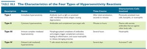
Hypersensitivity refers to an exaggerated immune response against a foreign antigen, resulting in tissue damage. There are four main types:
Type I (Immediate): IgE-mediated, rapid onset (allergies)
Type II (Cytotoxic): Antibody-mediated destruction of cells
Type III (Immune Complex-Mediated): Immune complex deposition and inflammation
Type IV (Delayed or Cell-Mediated): T cell-mediated, delayed response

Type I (Immediate) Hypersensitivity
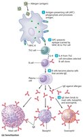
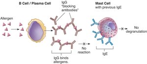
Type I hypersensitivity develops within minutes of exposure to an allergen and is commonly known as an allergy. It involves the production of IgE antibodies, which bind to mast cells, basophils, and eosinophils, leading to the release of inflammatory mediators upon re-exposure to the allergen.
Allergens: Antigens that trigger allergic reactions
Genetic predisposition: Susceptibility to allergies is often inherited
Hygiene hypothesis: Reduced early-life immune stimulation increases allergy risk
Mechanism of Type I Hypersensitivity
Sensitization: Initial exposure leads to IgE production
Degranulation: Re-exposure causes release of histamine and other mediators
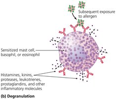
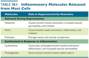
Inflammatory Molecules Released from Mast Cells
Molecule | Role in Hypersensitivity Reactions |
|---|---|
Histamine | Causes smooth muscle contraction, increased vascular permeability, and irritation |
Kinins | Cause smooth muscle contraction, inflammation, and irritation |
Proteases | Damage tissues and activate complement |
Leukotrienes | Cause slow, prolonged smooth muscle contraction, inflammation, and increased vascular permeability |
Prostaglandins | Some contract smooth muscle; others relax it |
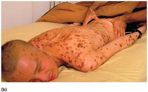
Clinical Manifestations
Localized reactions: Hay fever, asthma, hives (urticaria)
Systemic reactions: Anaphylactic shock, requiring immediate treatment with epinephrine

Diagnosis, Prevention, and Treatment
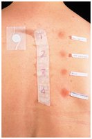
Diagnosis: Detection of allergen-specific IgE (e.g., ImmunoCAP test), skin testing
Prevention: Allergen avoidance, immunotherapy (allergy shots)
Treatment: Antihistamines, glucocorticoids, bronchodilators, epinephrine for severe reactions
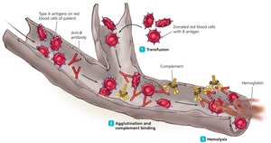
Type II (Cytotoxic) Hypersensitivity
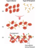
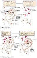
Type II hypersensitivity involves the destruction of cells by antibodies and complement. It is responsible for transfusion reactions and hemolytic disease of the newborn.
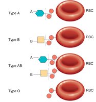
ABO blood group incompatibility: Transfusion of incompatible blood leads to hemolysis
Rh incompatibility: Rh-negative mothers may develop antibodies against Rh-positive fetal cells
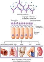
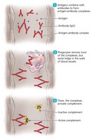
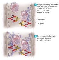
Type III (Immune Complex-Mediated) Hypersensitivity
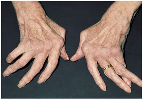
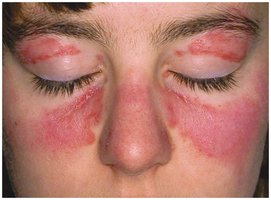
Type III hypersensitivity is caused by the formation of immune complexes that deposit in tissues, activating complement and causing inflammation and tissue damage. Examples include hypersensitivity pneumonitis, glomerulonephritis, rheumatoid arthritis, and systemic lupus erythematosus (SLE).
Localized reactions: Hypersensitivity pneumonitis, glomerulonephritis
Systemic reactions: Rheumatoid arthritis, SLE
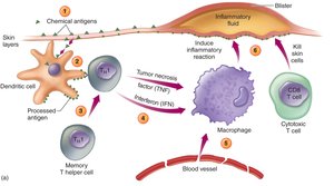
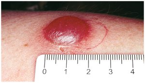
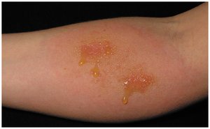
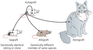
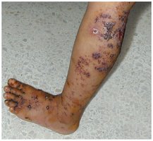
Type IV (Delayed or Cell-Mediated) Hypersensitivity
Type IV hypersensitivity is mediated by T cells and occurs 12–24 hours after antigen exposure. It includes contact dermatitis, the tuberculin skin test, and graft rejection.
Tuberculin test: Used to diagnose exposure to Mycobacterium tuberculosis
Contact dermatitis: Skin rash caused by contact with allergens (e.g., poison ivy, latex)
Graft rejection: Immune response against transplanted tissues or organs
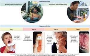
Autoimmune Diseases
Overview and Causes
Autoimmune diseases occur when the immune system attacks the body's own tissues due to a breakdown in self-tolerance. They can be systemic or organ-specific and are influenced by genetic, hormonal, and environmental factors.
Systemic: Affect multiple organs (e.g., SLE)
Single-organ: Target specific organs (e.g., type 1 diabetes, Graves disease, multiple sclerosis)
Examples
Autoimmune hemolytic anemia: Antibodies against red blood cells cause anemia
Type 1 diabetes mellitus: Immune destruction of pancreatic islet cells
Graves disease: Antibodies stimulate thyroid hormone production
Multiple sclerosis: Cytotoxic T cells destroy myelin in the nervous system
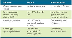
Immunodeficiency Diseases
Types and Causes
Immunodeficiency diseases result from defective immune mechanisms and are classified as primary (congenital) or secondary (acquired).
Primary immunodeficiencies: Genetic or developmental defects present from birth
Secondary immunodeficiencies: Result from external factors such as infections (e.g., HIV), malnutrition, or immunosuppressive drugs
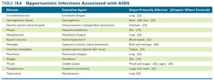
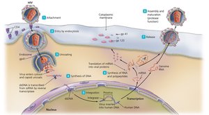
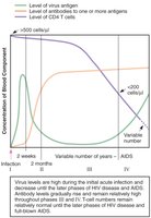
Acquired Immunodeficiency Syndrome (AIDS)
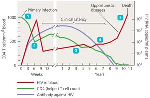
AIDS is caused by infection with the human immunodeficiency virus (HIV), which targets CD4+ T cells, leading to severe immune suppression and susceptibility to opportunistic infections and cancers.
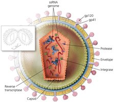
HIV characteristics: Enveloped, +ssRNA retrovirus; uses reverse transcriptase
Transmission: Blood, semen, vaginal secretions, breast milk
Diagnosis: Detection of HIV antibodies (ELISA, Western blot), low CD4+ T cell count, presence of opportunistic infections
Treatment: Antiretroviral therapy (ART) reduces viral replication but does not cure infection
Prevention: Safe sex, clean needles, screening blood products, pre-exposure prophylaxis

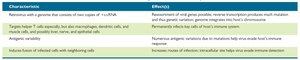
Summary Table: Characteristics of Hypersensitivity Types
Type | Name | Cause | Time Course | Characteristic Cells |
|---|---|---|---|---|
I | Immediate hypersensitivity | IgE against soluble antigen | After initial sensitization, seconds to minutes | Mast cells, basophils, eosinophils |
II | Cytotoxic hypersensitivity | IgG or IgM against cell surface antigen | Minutes to hours | Phagocytes, NK cells |
III | Immune complex-mediated hypersensitivity | Immune complexes of antibody and antigen | Several hours | Neutrophils |
IV | Delayed (cell-mediated) hypersensitivity | T cells attack body cells | Several days | Activated T cells |