 Back
BackImmune Disorders: Hypersensitivity and Autoimmunity
Study Guide - Smart Notes
 Tailored notes based on your materials, expanded with key definitions, examples, and context.
Immune Disorders
Overview of Hypersensitivity and Autoimmunity
Immune disorders arise when the immune system responds inappropriately, either by overreacting to harmless antigens (hypersensitivity) or by attacking the body’s own tissues (autoimmunity). Understanding these responses is crucial for diagnosing and treating related diseases.
Types of Hypersensitivity Reactions
Classification and Key Characteristics
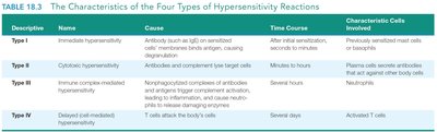
Hypersensitivity reactions are classified into four types based on their mechanisms, timing, and cellular mediators. Each type is associated with distinct clinical manifestations and underlying immunological processes.
Descriptive | Name | Cause | Time Course | Characteristic Cells Involved |
|---|---|---|---|---|
Type I | Immediate hypersensitivity | Antibody (mainly IgE) on sensitized cells interacts with antigen, causing degranulation | After initial sensitization, seconds to minutes | Previously sensitized mast cells or basophils |
Type II | Cytotoxic hypersensitivity | Antibodies and complement lyse target cells | Minutes to hours | Plasma cells secrete antibodies that act against other body cells |
Type III | Immune complex–mediated hypersensitivity | Nonphagocytized complexes of antibody and antigen trigger complement activation, leading to inflammatory damage | Several hours | Neutrophils |
Type IV | Delayed (cell-mediated) hypersensitivity | T cells attack the body’s cells | Several days | Activated T cells |
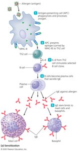
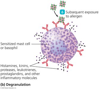
Type I (Immediate) Hypersensitivity
Type I hypersensitivity, also known as immediate or anaphylactic hypersensitivity, is characterized by a rapid allergic reaction following exposure to an allergen. This response is mediated by IgE antibodies and involves mast cell or basophil degranulation, releasing inflammatory mediators such as histamine.
Key Features: Rapid onset (seconds to minutes), involves allergens, and can be localized (e.g., hay fever) or systemic (e.g., anaphylaxis).
Mechanism: Sensitization occurs upon first exposure; subsequent exposures trigger degranulation and mediator release.
Examples: Allergic rhinitis, asthma, food allergies, anaphylactic shock.
Type II (Cytotoxic) Hypersensitivity
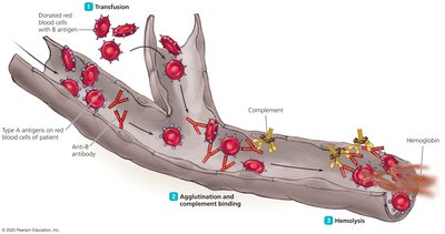
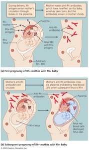
Type II hypersensitivity involves the destruction of cells by antibodies (IgG or IgM) and complement. This reaction is often seen in blood transfusion incompatibilities and certain autoimmune diseases.
Key Features: Antibody-mediated cell destruction, complement activation, and phagocytosis.
Examples: Hemolytic transfusion reactions, hemolytic disease of the newborn (Rh incompatibility).
Type III (Immune Complex–Mediated) Hypersensitivity
Type III hypersensitivity is caused by the formation of immune complexes (antigen-antibody complexes) that are not efficiently cleared. These complexes deposit in tissues, triggering inflammation and tissue damage.
Key Features: Immune complex deposition, complement activation, neutrophil recruitment.
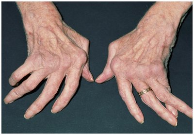
Examples: Systemic lupus erythematosus (SLE), rheumatoid arthritis (RA), glomerulonephritis.
Type IV (Delayed or Cell-Mediated) Hypersensitivity
Type IV hypersensitivity is mediated by T cells rather than antibodies. The reaction develops over 12–24 hours and involves macrophage and T cell recruitment to the site of antigen exposure.
Key Features: Delayed onset, T cell-mediated, tissue damage due to cellular immune response.
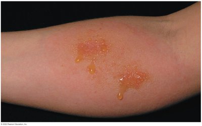
Examples: Tuberculin skin test, contact dermatitis (e.g., poison ivy), graft rejection.
Autoimmune Diseases
Overview and Causes
Autoimmune diseases occur when the immune system mistakenly targets self-antigens, leading to tissue damage and chronic inflammation. The exact causes are multifactorial, involving genetic, hormonal, and environmental factors.
Key Features: Loss of self-tolerance, production of autoantibodies or autoreactive T cells, chronic inflammation.
Examples: Type I diabetes, rheumatoid arthritis, multiple sclerosis, lupus.
Hypotheses for the Causes of Autoimmunity
Several hypotheses have been proposed to explain the development of autoimmune diseases:
Estrogen may stimulate destruction of tissue by cytotoxic T cells.
Maternal or fetal cells crossing the placenta may trigger autoimmunity.
Environmental factors such as viral infections can initiate autoimmunity.
Genetic predisposition, especially certain MHC genes.
T cells may encounter self-antigens that are normally hidden.
Molecular mimicry by microorganisms may trigger autoimmunity.
Failure of normal immune regulation mechanisms.
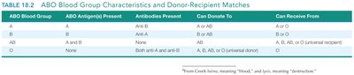
Summary Table: ABO Blood Group Compatibility
ABO Blood Group | ABO Antigen(s) Present | Antibodies Present | Can Donate To | Can Receive From |
|---|---|---|---|---|
A | A | Anti-B | A or AB | A or O |
B | B | Anti-A | B or AB | B or O |
AB | A and B | None | AB only | A, B, AB, or O (universal recipient) |
O | None | Both anti-A and anti-B | A, B, AB, or O (universal donor) | O only |
Key Points for Exam Preparation
Know the four types of hypersensitivity, their mechanisms, and clinical examples.
Understand the immunological basis of autoimmune diseases and the main hypotheses for their causes.
Be able to interpret blood group compatibility and the consequences of mismatched transfusions.
