 Back
BackImmune System Disorders: Hypersensitivity, Autoimmunity, and Immunodeficiency
Study Guide - Smart Notes
 Tailored notes based on your materials, expanded with key definitions, examples, and context.
Immune System Disorders
Overview
Immune system disorders arise when the immune response is either excessive, misdirected, or insufficient. These disorders include hypersensitivity reactions (allergies), autoimmune diseases, and immunodeficiency syndromes. Understanding these conditions is crucial for recognizing how the immune system can contribute to disease as well as protection.
Hypersensitivity Reactions
Definition and General Concepts
Hypersensitivity refers to an antigenic response that exceeds normal reactions, often resulting in tissue damage.
The antigen that triggers hypersensitivity is called an allergen.
There are four main types of hypersensitivity reactions, classified by their mechanisms and timing.
Type I Hypersensitivity (Anaphylactic Response)
This type involves an immediate allergic reaction mediated by IgE antibodies. It is the classic mechanism behind allergies such as hay fever, asthma, and anaphylactic shock.
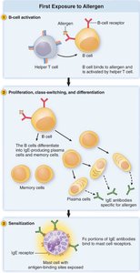
First Exposure: B cells recognize the allergen and, with help from T cells, differentiate into plasma cells that produce IgE antibodies. These IgE molecules bind to mast cells and basophils, sensitizing them for future encounters.
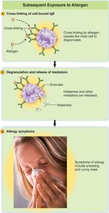
Second Exposure: The allergen cross-links IgE on mast cells or basophils, triggering the release of histamine and other mediators.
Symptoms: Vasodilation, redness, swelling, itchiness, increased mucous production, and bronchial constriction. Systemic exposure can cause anaphylactic shock (sudden drop in blood pressure).
Example: Peanut allergy.
Treatments for Type I Hypersensitivity
Antihistamines: Block histamine receptors, reducing symptoms such as itching and swelling. Effective for mild allergies (e.g., hay fever).
Epinephrine: Used for anaphylactic shock; acts as a vasoconstrictor to increase blood pressure and counteract severe allergic reactions.
Allergy Shots (Immunotherapy): Gradual exposure to increasing doses of allergen induces IgG production, which neutralizes the allergen before it can bind IgE, preventing histamine release.
Type II Hypersensitivity (Antibody-Dependent Cytotoxicity)
This reaction involves antibodies (IgG or IgM) directed against antigens on the surface of cells, leading to cell destruction via complement activation or phagocytosis. Commonly seen in blood transfusion reactions and hemolytic disease of the newborn.
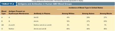
ABO Blood Group System: Antibodies target A or B antigens on red blood cells, causing cell lysis if mismatched blood is transfused.
Blood Type | Antigen Present | Antibody in Plasma | Incidence (Whites) | Incidence (Asians) | Incidence (Blacks) |
|---|---|---|---|---|---|
A | A | Anti-B | 41% | 28% | 27% |
B | B | Anti-A | 10% | 27% | 20% |
AB | A and B | Neither anti-A nor anti-B | 4% | 5% | 7% |
O | Neither | Anti-A and anti-B | 45% | 40% | 46% |
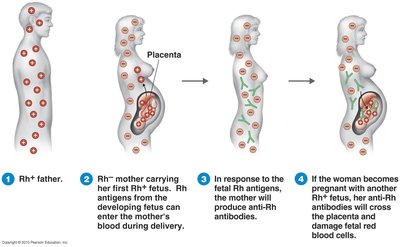
Rh Factor: An antigen on red blood cells. If an Rh- mother carries an Rh+ fetus, she may develop anti-Rh antibodies. In subsequent pregnancies, these antibodies can cross the placenta and destroy fetal red blood cells, causing hemolytic disease of the newborn.
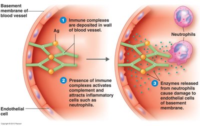
Type III Hypersensitivity (Immune Complex-Mediated)
Occurs when antigen-antibody complexes form in the bloodstream and deposit in tissues, leading to inflammation and tissue damage. This can activate complement and attract neutrophils, which release enzymes that damage tissues.
Mechanism: Immune complexes become trapped in membranes (e.g., blood vessels), activating complement and inflammatory cells.
Result: Inflammation and tissue injury, such as vasculitis or glomerulonephritis.
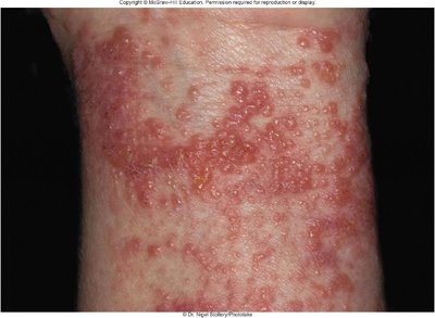
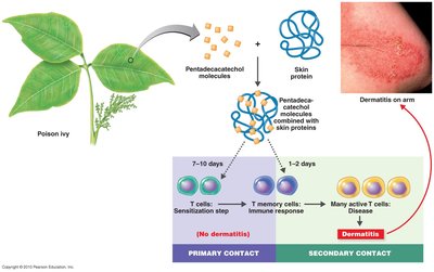
Type IV Hypersensitivity (Delayed-Type, Cell-Mediated)
This reaction is mediated by T cells rather than antibodies and typically occurs 24–72 hours after exposure to the antigen. It is responsible for contact dermatitis and some transplant rejection reactions.
Mechanism: Small chemicals (haptens) bind to skin proteins, forming new antigens that activate memory T cells. Upon re-exposure, cytotoxic T cells attack the altered skin cells, causing localized inflammation.
Symptoms: Red, itchy, swollen skin (e.g., poison ivy dermatitis).
Transplantation and Immune Response
Transplant Rejection and Graft-vs-Host Disease
Transplantation: Involves immune responses against non-self MHC molecules on transplanted tissues, primarily mediated by cytotoxic T cells (Tc) and natural killer (NK) cells.
Prevention: Matching donor and recipient tissues and using immunosuppressive drugs (e.g., cyclosporin) to prevent T cell activation.
Graft-vs-Host Disease: Donor T cells attack recipient tissues, especially after bone marrow transplants.
Risks: Immunosuppressed patients are highly susceptible to infections and must be kept in isolation.
Immunodeficiency Disorders
Primary (Congenital) Immunodeficiency
Genetic defects resulting in non-functional T and/or B cells.
Example: Severe Combined Immunodeficiency Disease (SCID) leads to extreme vulnerability to infections.
Acquired Immunodeficiency
Develops after birth due to infections or other factors.
Example: Acquired Immunodeficiency Syndrome (AIDS) is caused by HIV, which destroys T helper cells, macrophages, and dendritic cells, severely impairing both antibody and cell-mediated immunity.
Autoimmune Diseases
Overview
Autoimmune diseases occur when the immune system mistakenly attacks the body's own tissues. Causes may include infections, genetic predisposition, or unknown factors.
Rheumatic Fever: Follows Streptococcus pyogenes infection; antibodies against bacterial M protein cross-react with heart tissue, causing damage.
Rheumatoid Arthritis: IgM, IgG, and complement attack collagen in joints, leading to chronic inflammation and pain.
Lupus (Systemic Lupus Erythematosus): Antibodies target chromatin (DNA-protein complexes), forming immune complexes that deposit in joints, kidneys, and blood vessels.
Multiple Sclerosis: T cells and macrophages attack the myelin sheath of neurons, causing neurological symptoms. May be triggered by Epstein-Barr virus infection.
