 Back
BackInfection, Infectious Disease, and Epidemiology – Study Notes
Study Guide - Smart Notes
 Tailored notes based on your materials, expanded with key definitions, examples, and context.
Chapter 14: Infection, Infectious Disease, and Epidemiology
Introduction to Pathology and Infectious Disease
Pathology is the study of disease, including its causes (etiology), development (pathogenesis), and effects on the body. Infectious diseases are caused by pathogenic microorganisms that colonize and impair normal body function. Epidemiology is the study of the occurrence, distribution, and spread of diseases in populations.
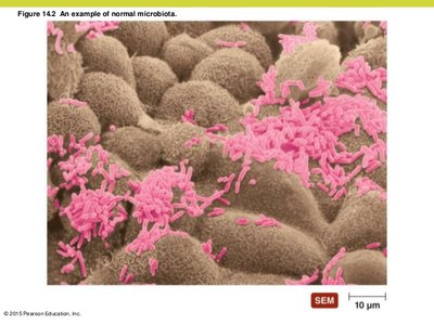
Normal Microbiota and Symbiosis
Resident and Transient Flora
Resident flora (normal microbiota): Microorganisms that remain a part of the microbiome for most of a person’s life. They are typically commensal, feeding on cellular wastes and dead cells without causing harm.
Transient flora: Microbes that remain in the body for only a short period (hours to months) and are found in the same locations as resident flora. They cannot persist due to competition, elimination by body defenses, or environmental changes.
Symbiotic Relationships
Mutualism: Both organisms benefit.
Commensalism: One organism benefits, the other is unaffected.
Amensalism: One organism is harmed, the other is unaffected.
Parasitism: One organism benefits at the expense of the other.
Acquisition and Role of Normal Microbiota
Initial acquisition occurs during birth and the first months of life.
Normal microbiota protect the host by occupying niches, competing for nutrients, and producing substances harmful to pathogens (e.g., acids, bacteriocins).
Opportunistic Pathogens
Normal microbiota can become opportunistic pathogens under certain conditions, such as:
Immune suppression (e.g., HIV, chemotherapy)
Changes in the normal microbiota (e.g., antibiotics, diet)
Introduction into unusual body sites
Stressful conditions (e.g., hormonal changes, emotional stress)
Sterile Areas of the Body
Internal organs (heart, kidneys, liver), muscle, connective tissue, blood, urine (as produced in kidneys), cerebrospinal fluid are normally sterile.
Areas open to the environment (skin, GI tract, upper respiratory tract, distal urogenital tract) are colonized by microbiota.
Reservoirs and Transmission of Infectious Diseases
Reservoirs of Infection
Reservoirs are sites where pathogens are maintained as sources of infection:
Animal reservoirs: Zoonoses are diseases naturally spread from animals to humans (e.g., direct contact, eating animals, arthropod vectors).
Human carriers: Asymptomatic individuals who can transmit pathogens (e.g., Mary Mallon, "Typhoid Mary").
Nonliving reservoirs: Soil, water, and food contaminated by feces or urine.
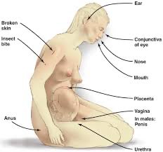
Portals of Entry
Pathogens enter the body through specific portals:
Skin: Through cuts, abrasions, bites, or by burrowing (e.g., parasitic larvae).
Mucous membranes: Line body cavities open to the environment; common entry points due to their thin, moist nature.
Placenta: Some pathogens can cross the placenta during pregnancy (e.g., Toxoplasma).
Parenteral route: Pathogens deposited directly into tissues (e.g., via needles, catheters).
Virulence Factors and Pathogenesis
Virulence and Pathogenicity
Pathogenicity: The ability of a microorganism to cause disease.
Virulence: The degree of pathogenicity, determined by factors such as adhesion, biofilm formation, extracellular enzymes, toxins, and antiphagocytic factors.
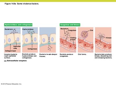
Extracellular Enzymes
Secreted by pathogens to dissolve structural chemicals and facilitate invasion (e.g., hyaluronidase, collagenase, coagulase, kinase).
Loss of these enzymes reduces virulence.
Toxins
Exotoxins: Proteins secreted by pathogens (e.g., cholera toxin, botulinum toxin, tetanus toxin). Highly toxic and specific in action.
Endotoxins: Lipopolysaccharides (LPS) from the outer membrane of Gram-negative bacteria, released upon cell death. Lipid A component is toxic and can cause toxic shock.
Feature | Exotoxin | Endotoxin |
|---|---|---|
Source | Gram-positive and Gram-negative bacteria | Gram-negative bacteria only |
Chemical Nature | Protein | Lipopolysaccharide (LPS) |
Heat Stability | Unstable (destroyed by heat) | Stable |
Effect | Specific for target cells | Generalized (fever, shock) |
Toxicity | High | Low (but can be fatal in large amounts) |
Antiphagocytic Factors
Bacterial capsules and antiphagocytic enzymes help pathogens evade host immune responses.
Examples: Capsules prevent phagocytosis; enzymes prevent lysosome fusion or kill phagocytes.
Development and Stages of Infectious Disease
Predisposing Factors
Short urethra in females, inherited traits, climate, fatigue, age, lifestyle, nutrition, chemotherapy can increase susceptibility.
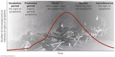
Stages of Disease
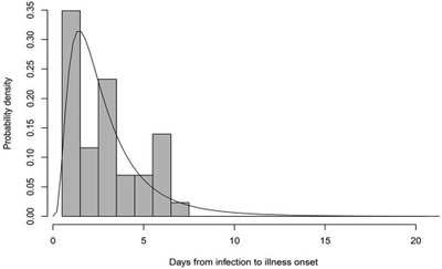
Incubation period: Time between infection and first symptoms.
Prodromal period: Early, mild symptoms.
Period of illness: Most severe symptoms.
Period of decline: Symptoms subside.
Period of convalescence: Recovery and return to normal.
Transmission of Infectious Diseases
Portals of Exit
Pathogens exit via skin, mucous membranes, saliva, respiratory droplets, urine, feces, etc.
Modes of Transmission
Direct contact: Person-to-person (e.g., touching, kissing).
Indirect contact: Via fomites (e.g., needles, toys).
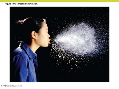
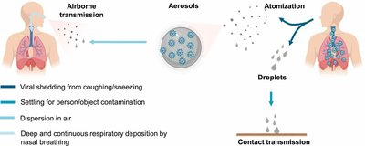
Droplet transmission: Via droplets expelled during coughing or sneezing, traveling less than 1 meter.
Vehicle Transmission
Airborne: Pathogens travel more than 1 meter in aerosols (e.g., tuberculosis).
Waterborne: Contaminated water spreads gastrointestinal diseases (e.g., cholera).
Foodborne: Ingestion of contaminated, undercooked, or unrefrigerated food.
Vector Transmission
Biological vectors: Arthropods (e.g., mosquitoes, ticks) that serve as hosts and transmit pathogens.
Mechanical vectors: Arthropods (e.g., flies, cockroaches) that passively carry pathogens on their bodies.
Classification of Infectious Diseases
Communicable, Contagious, and Noncommunicable Diseases
Communicable: Spread from host to host (e.g., typhoid fever, tuberculosis).
Contagious: Easily spread (e.g., measles, influenza).
Noncommunicable: Not spread between hosts (e.g., tetanus, toxic shock).
Types of Infections
Local infection: Limited to a small area (e.g., boils).
Systemic infection: Spread throughout the body (e.g., gangrene).
Focal infection: Starts local, spreads systemically (e.g., streptococcal infections).
Sepsis: Toxic inflammatory condition from spread of microbes or toxins.
Bacteremia: Bacteria in the blood.
Septicemia: Growth of bacteria in the blood.
Toxemia: Toxins in the bloodstream.
Occurrence of Disease
Endemic: Constantly present in a population (e.g., common cold).
Sporadic: Occurs occasionally (e.g., Creutzfeldt-Jakob disease).
Epidemic: Many hosts in a short time (e.g., polio).
Pandemic: Worldwide epidemic (e.g., AIDS, COVID-19).
Herd immunity: Immunity in most of the population reduces disease spread.
Epidemiology and Public Health
Measures of Disease Frequency
Incidence: Number of new cases in a given area during a specific period.
Prevalence: Total number of cases (new and existing) in a given area during a specific period.
Descriptive, Analytical, and Experimental Epidemiology
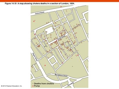
Descriptive epidemiology: Tabulation of data, identification of index case (first documented case), and mapping outbreaks (e.g., John Snow’s cholera map).
Analytical epidemiology: Determines probable cause, mode of transmission, and prevention methods.
Experimental epidemiology: Tests hypotheses about disease prevention and treatment.
Notable Figures in Epidemiology and Infection Control
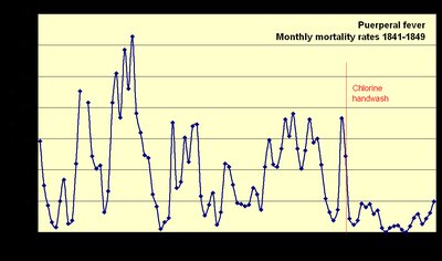
Ignaz Semmelweis: Demonstrated handwashing reduces puerperal fever in hospitals.
Joseph Lister: Introduced surgical antisepsis using carbolic acid (phenol).
John Snow: Mapped cholera cases, identified contaminated water as the source.
Mary Mallon (Typhoid Mary): Asymptomatic carrier of Salmonella typhi.
Florence Nightingale: Improved hospital sanitation, reduced infection rates.
Public Health Organizations
US Centers for Disease Control and Prevention (CDC): Tracks disease occurrence, causes, transmission, and prevention in the US.
World Health Organization (WHO): Coordinates international public health efforts.
Summary Table: Key Terms and Concepts
Term | Definition |
|---|---|
Pathology | Study of disease |
Etiology | Study of the cause of disease |
Pathogenesis | Development of disease |
Infection | Colonization of the body by pathogens |
Disease | Abnormal state in which the body is not functioning normally |
Incidence | Number of new cases in a given period |
Prevalence | Total number of cases in a given period |
Additional info: These notes integrate textbook content, lecture points, and relevant images to provide a comprehensive overview of infection, infectious disease, and epidemiology for microbiology students.
