Back
BackInfectious Diseases Affecting the Respiratory System: Microbiology Study Notes
Study Guide - Smart Notes
 Tailored notes based on your materials, expanded with key definitions, examples, and context.
Respiratory System Anatomy and Defenses
Anatomical Features of the Respiratory Tract
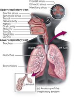
The respiratory tract is divided into upper and lower sections, each with distinct anatomical structures and functions. Understanding these divisions is essential for recognizing how pathogens invade and cause disease.
Upper respiratory tract: Includes the mouth, nose, nasal cavity, sinuses, throat (pharynx), epiglottis, and larynx.
Lower respiratory tract: Comprises the trachea, bronchi, bronchioles, and alveoli, where gas exchange occurs.
Natural Defenses of the Respiratory Tract
The respiratory tract is equipped with multiple defense mechanisms to prevent infection by inhaled pathogens.
Nasal hair: Traps large particles and pathogens.
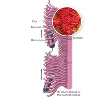
Cilia: Propel trapped particles upward and out of the tract (mucociliary escalator).
Mucus: Acts as a sticky barrier to trap microorganisms.
Immune defenses: Include complement proteins, antimicrobial peptides, cytokines, macrophages, and secretory IgA.
Normal Biota of the Respiratory Tract
Composition and Role of Normal Biota
The upper respiratory tract harbors a diverse community of commensal microorganisms, while even the lungs contain a limited normal biota. These organisms play a crucial role in maintaining respiratory health.
Common normal biota: Streptococcus pyogenes, Haemophilus influenzae, Streptococcus pneumoniae, Neisseria meningitidis, Staphylococcus aureus, and fungi such as Candida albicans.
Microbial antagonism: Normal biota compete with pathogens for resources and space, reducing the risk of infection (e.g., Lactobacillus sakei suppresses Corynebacterium tuberculostearicum in the sinuses).
Biota variation: The composition of the lung microbiome differs in patients with chronic diseases (COPD, asthma, cystic fibrosis) and between smokers and nonsmokers.
Upper Respiratory Tract Diseases
Pharyngitis
Pharyngitis is inflammation of the throat, often presenting with pain, redness, and swelling. Bacterial pharyngitis is typically more severe than viral forms and may be accompanied by fever, headache, and nausea.
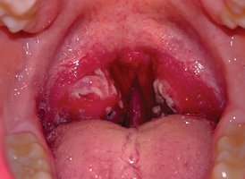
Major causative agents: Viruses (common cold viruses), Streptococcus pyogenes (Group A Streptococcus), and Fusobacterium necrophorum.
Complications: Untreated S. pyogenes can lead to scarlet fever (erythrogenic toxin, sandpaper-like rash, high fever) and rheumatic fever (autoimmune reaction affecting the heart).
Streptococcus pyogenes: Structure and Virulence
Streptococcus pyogenes is a Gram-positive coccus that grows in chains, forms capsules and slime layers, and is a facultative anaerobe. It does not produce catalase but survives using a peroxidase system.
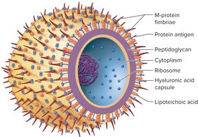
Virulence factors:
Surface antigens mimic host proteins (immune evasion).
Superantigens trigger excessive immune responses.
Specialized polysaccharides protect against lysozyme.
Lipoteichoic acid and M protein aid adherence and resist phagocytosis.
Hyaluronic acid capsule enhances adhesiveness.
Extracellular toxins: Streptolysins (O and S) cause beta-hemolysis; erythrogenic toxin (in lysogenic strains) causes rash and fever.
Diagnosis and Treatment of Streptococcal Pharyngitis
Diagnosis involves rapid antigen detection tests and culturing on sheep blood agar to observe beta-hemolysis. The bacitracin disc test helps distinguish S. pyogenes from other beta-hemolytic streptococci.

Prevention: No vaccine; hand hygiene is critical.
Treatment: Penicillin (first-line), cephalexin for penicillin-allergic patients. Antibiotics prevent serious sequelae.
Other Upper Respiratory Tract Infections
The Common Cold: Caused by over 200 viruses (rhinoviruses, coronaviruses, adenoviruses, RSV). Symptoms include sneezing, scratchy throat, and runny nose. Transmission is via droplets and indirect contact.
Sinusitis: Can be viral, bacterial, or fungal. Noninfectious causes include allergies and structural abnormalities. Treated with antibiotics (bacterial), antifungals/surgery (fungal).
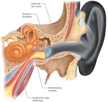
Acute Otitis Media (Ear Infection): Often follows viral upper respiratory infections. Common agents: Streptococcus pneumoniae, Candida auris. Prevention includes vaccines (e.g., Prevnar); treatment may involve antibiotics or tympanic membrane tubes.
Lower Respiratory Tract Diseases
Pneumonia
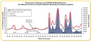
Pneumonia is an inflammatory condition where fluid fills the alveoli. It can be caused by bacteria, viruses, or fungi, and is a leading cause of death in children under five worldwide.
Community-acquired pneumonia: Common agents include Streptococcus pneumoniae (pneumococcus), Legionella pneumophila, Mycoplasma pneumoniae, Chlamydophila pneumoniae, Histoplasma capsulatum, hantavirus, and respiratory viruses.
Healthcare-associated pneumonia: Often associated with mechanical ventilation; common agents include MRSA, Klebsiella pneumoniae, Enterobacter, E. coli, Pseudomonas aeruginosa, and Acinetobacter.
Influenza
Influenza is a viral infection with cyclical outbreaks, especially in winter. It can cause pandemics due to the virus's genetic variability.
Causative agents: Influenza A, B, and C viruses (family Orthomyxoviridae).
Structure: Spherical, enveloped virus with hemagglutinin (H) and neuraminidase (N) spikes, ion channels, and a segmented ssRNA genome.
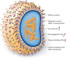
Antigenic drift: Gradual mutations in H and N proteins, requiring annual vaccine updates.
Antigenic shift: Major genetic reassortment between different influenza viruses, often in pigs, leading to new pandemic strains.
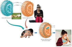
Transmission: Aerosols, droplets, and fomites; facilitated by crowding and dry air.
Diagnosis: RT-PCR is preferred; culture and non-culture tests are also used.
Prevention: Vaccination (inactivated or live attenuated); high-dose vaccine for older adults.
Treatment: Early antiviral drugs (zanamivir, oseltamivir, baloxavir).
Whooping Cough (Pertussis)
Caused by Bordetella pertussis, whooping cough is transmitted by droplets and progresses through catarrhal, paroxysmal, and convalescent stages. Prevention relies on the DTaP vaccine, but immunity is not lifelong.
Respiratory Syncytial Virus (RSV) Infection
RSV is a major cause of respiratory illness in infants and young children, especially in winter and early spring. It forms syncytia (fused cells) and is prevented by passive antibody administration in high-risk children and vaccines for older adults.
Tuberculosis
Overview and Clinical Forms
Tuberculosis (TB) is a chronic infectious disease caused by Mycobacterium tuberculosis. It primarily affects the lungs but can disseminate to other organs.
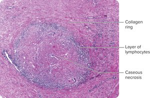
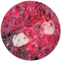
Primary TB: Infection is contained in the lungs, forming granulomas (tubercles) with caseous necrosis.
Extrapulmonary TB: Involves lymph nodes, kidneys, bones, genital tract, brain, and meninges; more common in immunosuppressed patients and children.
Secondary (reactivation) TB: Dormant bacteria reactivate, causing severe symptoms and high mortality if untreated.
Pathogenesis and Virulence
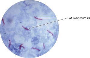
Mycobacterium tuberculosis is an acid-fast bacillus with a waxy cell wall (mycolic acids), enhancing survival in the environment and within macrophages. The cord factor is linked to virulence.
Transmission: Airborne droplets; bacteria can survive in aerosols for months.
Risk factors: Malnutrition, immune suppression, poor healthcare access, lung damage, and genetics.
Diagnosis, Prevention, and Treatment
Diagnosis: Tuberculin skin test (Mantoux), interferon-gamma release assays (IGRAs), gene amplification, antimicrobial susceptibility testing, and acid-fast staining.
Prevention: Isolation of active cases, BCG vaccine (not used in the US), limiting exposure to airborne particles.
Treatment: Long-term antibiotic regimens (4–9 months) with rifampin, rifapentine, and isoniazid. Noncompliance leads to multidrug-resistant TB (MDR-TB) and extensively drug-resistant TB (XDR-TB).
MDR-TB and XDR-TB
MDR-TB: Resistant to at least isoniazid and rifampin; requires prolonged treatment with multiple drugs; high mortality.
XDR-TB: MDR-TB strains with resistance to two additional drugs; very limited treatment options and extremely high mortality.
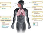
Summary Table: Major Respiratory Pathogens and Diseases
Pathogen Type | Diseases Caused | Examples |
|---|---|---|
Bacteria | Sinusitis, pneumonia, otitis media, pharyngitis, whooping cough, tuberculosis | Streptococcus pneumoniae, Streptococcus pyogenes, Bordetella pertussis, Mycobacterium tuberculosis |
Viruses | Respiratory syncytial virus infection, pharyngitis, rhinitis, influenza, pneumonia | RSV, influenza virus, rhinovirus, coronavirus |
Fungi | Sinusitis, pneumonia | Histoplasma capsulatum, Pneumocystis jiroveci |