 Back
BackInnate and Adaptive Immunity: Lines of Defense in the Human Body
Study Guide - Smart Notes
 Tailored notes based on your materials, expanded with key definitions, examples, and context.
Immunity: Lines of Defense
Overview of Immunity
Immunity refers to the organism's defense mechanisms against infections. The immune system is organized into three main lines of defense, each with distinct characteristics, specificity, and cellular players. These lines of defense are classified as innate (nonspecific) or adaptive (specific) immunity, with varying requirements for activation and memory formation.
First Line (Innate, Nonspecific): Physical, chemical, and genetic barriers that prevent pathogen entry.
Second Line (Innate, Nonspecific): Cellular and molecular responses such as inflammation, phagocytosis, and complement activation.
Third Line (Adaptive, Specific): Highly specific responses involving T cells, B cells, and antibodies, with immunological memory.
First Line of Defense: Physical, Chemical, and Genetic Barriers
Physical Barriers
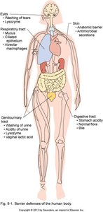
The first line of defense consists of anatomical and physiological barriers that block pathogen entry. These barriers are always present and active, providing immediate protection.
Skin: Multiple layers of tightly joined epithelial cells and dead keratinized cells form a tough barrier.
Mucous Membranes: Mucus traps microbes; cilia sweep them out of the respiratory tract.
Other Barriers: Nasal hair, ear wax, and normal body temperature contribute to defense.
Normal Flora: Commensal microbes compete for nutrients, alter pH, and stimulate antimicrobial compound production.
Washing and Removal Mechanisms
Continuous shedding of skin and mucosal cells
Flushing by sweat, tears, saliva, urine, and other secretions
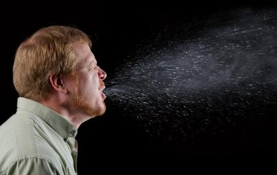
Mechanical actions: blinking, coughing, sneezing, vomiting, menstruation, ejaculation, and bleeding
Chemical Barriers
Chemical factors in body fluids and secretions inhibit or destroy pathogens.
Acidic pH: Stomach, vagina, and urine acidity inhibits microbial growth.
Salt and Acid in Sweat: Create an inhospitable environment for microbes.
Digestive Enzymes and Bile: Degrade microbial components in the intestines.
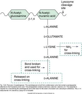
Lysozyme: Enzyme in tears, saliva, and other secretions that cleaves glycosidic bonds in peptidoglycan, weakening bacterial cell walls.
Lactoferrin and Transferrin: Bind iron, limiting its availability to microbes ("keep-away" strategy).
Antimicrobial Peptides: Defensins, histatins, cathelicidins, and others disrupt microbial membranes and inhibit biosynthesis.
Genetic Barriers
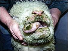
Some species and individuals possess genetic resistance to specific pathogens due to differences in biomolecule structure or the absence of cellular receptors required for infection.
Species Resistance: Inherent resistance to diseases affecting other species.
Individual Resistance: Genetic mutations or absence of receptors can confer immunity to certain infections.
Second and Third Lines of Defense: Innate and Adaptive Immunity
Activation and Interaction
If pathogens breach the first line, the second (innate) and third (adaptive) lines of defense are activated. These involve a coordinated response by immune cells, proteins, and signaling molecules. The innate response is rapid (minutes to hours), while the adaptive response takes longer (days to weeks) and requires activation by the innate system.
Recognition: Detection of danger signals or foreign molecules (MAMPs/DAMPs).
Activation: Recruitment and activation of immune cells and proteins.
Effector Phase: Elimination of pathogens and resolution of infection.
Lymphoid Organs and Immune Cell Development
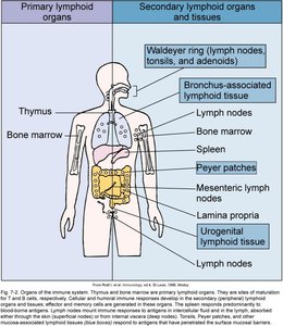
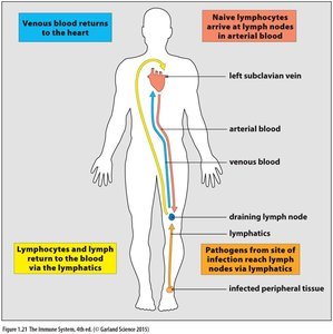
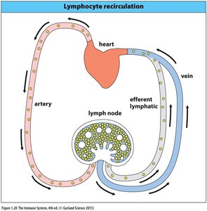
Immune cells develop and mature in primary lymphoid organs and are activated in secondary lymphoid organs. The lymphatic system facilitates immune cell circulation and pathogen detection.
Primary Lymphoid Organs: Bone marrow (hematopoiesis), thymus (T cell maturation)
Secondary Lymphoid Organs: Lymph nodes, spleen, mucosal-associated lymphoid tissue (MALT, GALT, BALT), Peyer's patches
Hematopoiesis and Immune Cell Lineages
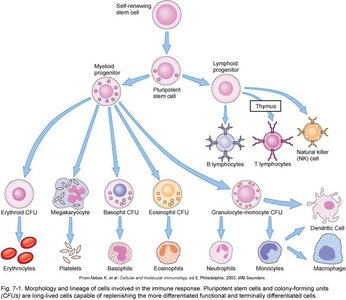
All immune cells originate from hematopoietic stem cells in the bone marrow, differentiating into myeloid and lymphoid lineages. These give rise to granulocytes, monocytes, macrophages, dendritic cells, lymphocytes, and other immune cells.
Innate Immune Cells
Granulocytes
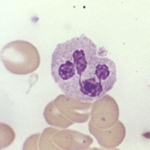
Neutrophils: Most abundant WBCs; phagocytic; multi-lobed nuclei; first responders to infection.
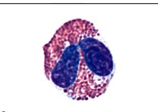
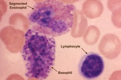
Eosinophils: Bilobed nuclei; combat eukaryotic parasites; weak phagocytes.
Basophils: Induce inflammation; granules stain blue; rare in circulation.
Mast Cells: Tissue-resident equivalents of basophils; mediate allergic responses.
Agranulocytes
Lymphocytes: B cells and T cells (adaptive immunity), Natural Killer (NK) cells (innate immunity).
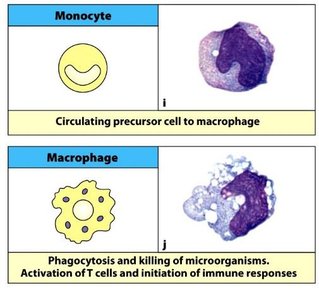
Monocytes: Circulating precursors to macrophages; kidney-shaped nucleus.
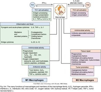
Macrophages: Mature from monocytes; phagocytose pathogens and present antigens.
Dendritic Cells: Trap pathogens and initiate immune responses; some derive from monocytes.
Cell Surface Markers (CD Markers)
Cluster of Differentiation (CD) markers are surface proteins used to identify and classify immune cells. Examples include CD3 (all T cells), CD4 (helper T cells, monocytes, macrophages, dendritic cells), and CD8 (cytotoxic T cells).
Phagocytosis and Extracellular Killing
Phagocytosis
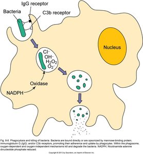
Phagocytes (neutrophils, monocytes, macrophages, dendritic cells) survey tissues, ingest, and destroy microbes and debris. Some phagocytes also activate adaptive immune responses.
Phagosome Formation: Engulfed material is enclosed in a phagosome, which fuses with a lysosome to form a phagolysosome for degradation.
Destruction Mechanisms: Reactive oxygen species, acids, antimicrobial peptides, and enzymes (e.g., proteases).
Extracellular Killing
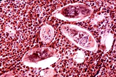
Eosinophils: Release lysosomal contents onto parasites (e.g., helminths).
Neutrophils: Release reactive oxygen species and form neutrophil extracellular traps (NETs).
Natural Killer (NK) Cells: Recognize and kill abnormal host cells (e.g., virally infected or tumor cells) by releasing perforins and granzymes.
Type I Interferons and Antiviral Defense
Type I Interferons (IFN-α, IFN-β)
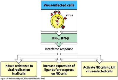
Produced by virally infected cells and certain dendritic cells, type I interferons bind to receptors on neighboring cells, inducing an antiviral state by upregulating hundreds of genes. They also enhance MHC class I expression and activate dendritic cells, macrophages, and NK cells.
Pattern Recognition and Immune Activation
Pattern Recognition Receptors (PRRs)
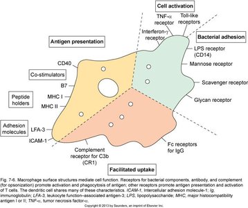
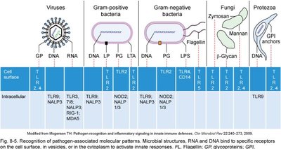
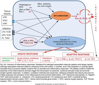
PRRs on host cells recognize conserved microbial motifs (MAMPs) or host-derived damage signals (DAMPs), triggering immune responses. Examples include Toll-like receptors (TLRs) and NOD-like receptors (NLRs).
Complement System
Complement Cascade
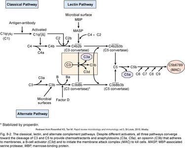
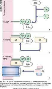
The complement system consists of 26 serum proteins that act in a cascade to tag or destroy microorganisms. There are three activation pathways:
Classical Pathway: Triggered by antibodies or PRR-bound MAMPs.
Lectin Pathway: Initiated by serum PRRs binding specific sugars on microbes.
Alternative Pathway: Activated by direct binding of complement proteins to microbial surfaces.
Major Complement Functions
C3b: Opsonization (enhances phagocytosis).
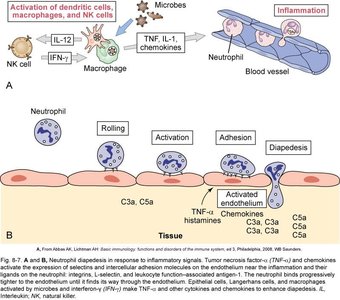
C3a and C5a: Promote inflammation and chemotaxis.
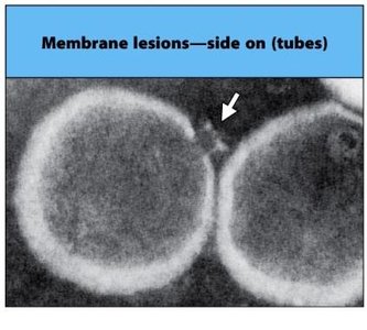
Membrane Attack Complex (MAC): Direct killing of Gram-negative bacteria and some virally infected cells.
Inflammatory Response
Characteristics and Signs
Inflammation is a general response to tissue damage or infection, characterized by redness (rubor), warmth (calor), swelling (tumor), pain (dolor), and sometimes loss of function. It involves increased blood flow, vascular permeability, and recruitment of immune cells.
Redness and Warmth: Due to increased circulation and vasodilation.
Swelling: Fluid accumulation (edema) from leaky blood vessels.
Pain: Stimulation of nerve endings by inflammatory mediators.
Loss of Function: May occur in severe inflammation.
Summary Table: Lines of Immune Defense
Line of Defense | Type | Specificity | Memory | Main Components |
|---|---|---|---|---|
First | Innate | Nonspecific | No | Physical, chemical, genetic barriers; normal flora |
Second | Innate | Nonspecific | Little | Phagocytes, inflammation, complement, NK cells, cytokines, fever |
Third | Adaptive | Specific | Yes | T cells, B cells, antibodies |
Additional info: The immune system's ability to distinguish self from non-self and to remember previous encounters with pathogens is fundamental to adaptive immunity. The interplay between innate and adaptive responses ensures effective defense and long-term protection.
