 Back
BackInnate Immunity: Nonspecific Defenses of the Host
Study Guide - Smart Notes
 Tailored notes based on your materials, expanded with key definitions, examples, and context.
Innate Immunity: Nonspecific Defenses of the Host
Overview of Immunity
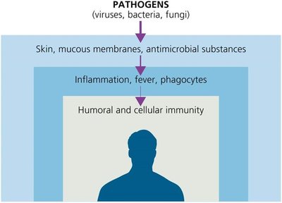
Immunity refers to the body's ability to ward off disease, while susceptibility is the lack of resistance to disease. The immune system is divided into innate (nonspecific) and adaptive (specific) immunity. Innate immunity provides immediate, general defense against pathogens and is present at birth, whereas adaptive immunity targets specific pathogens and develops memory for future responses.
Innate Immunity: Rapid, present at birth, acts against any pathogen.
Adaptive Immunity: Slower, specific to particular pathogens, has memory.
White Blood Cell (WBC) Counts: Used to assess immune status; high counts may indicate infection or autoimmune disease, while low counts may suggest viral infection or severe bacterial infection.
The Concept of Innate Immunity
Cells of the innate immune system recognize pathogens using pattern recognition receptors such as Toll-like receptors (TLRs), which bind to pathogen-associated molecular patterns (PAMPs) like LPS, flagellin, and peptidoglycan. This interaction triggers the release of cytokines, which regulate immune responses and recruit other immune cells.
PAMPs: Conserved microbial structures recognized by the immune system.
Cytokines: Signaling proteins that modulate immune responses.
First Line of Defense: Physical, Chemical, and Microbiological Barriers
Physical Factors
The first line of defense includes physical barriers that prevent pathogen entry.
Skin: Composed of the dermis (connective tissue) and epidermis (keratinized epithelial cells). Shedding and dryness inhibit microbial growth.
Mucous Membranes: Line the gastrointestinal, respiratory, and genitourinary tracts. Mucus traps microbes; ciliary escalator moves them out of the lungs.
Other Mechanisms: Lacrimal apparatus (tears), earwax, urine flow, vaginal secretions, peristalsis, defecation, vomiting, and diarrhea help remove microbes.
Chemical Factors
Chemical barriers further inhibit microbial growth.
Sebum: Forms a protective film and lowers skin pH (3–5).
Lysozyme: Enzyme in tears, saliva, and other secretions that destroys bacterial cell walls.
Low pH: Gastric juice (pH 1.2–3.0) and vaginal secretions (pH 3–5) inhibit or destroy microbes.
Normal Microbiota and Innate Immunity
Normal microbiota protect the host by competing with pathogens for nutrients and space, producing substances harmful to pathogens, and altering environmental conditions. They also play a role in immune system development.
Microbial Antagonism: Competition between normal microbiota and pathogens.
Commensalism: One organism benefits, the other is unharmed.
Opportunistic Pathogens: Normal microbiota that can cause disease under certain conditions (e.g., E. coli, S. aureus).
Probiotics: Live microbes administered for health benefits.
Prebiotics: Nutrients that promote growth of beneficial microbes.
Second Line of Defense: Cellular and Chemical Responses
Formed Elements in Blood
Blood contains cells and cell fragments suspended in plasma, including erythrocytes (RBCs), leukocytes (WBCs), and platelets. These are produced in red bone marrow through hematopoiesis.
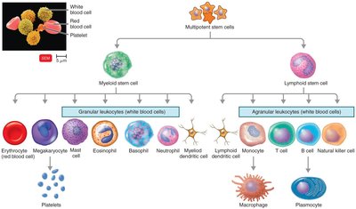
Leukocytes (White Blood Cells)
Leukocytes are divided into granulocytes and agranulocytes, each with specific roles in immunity.
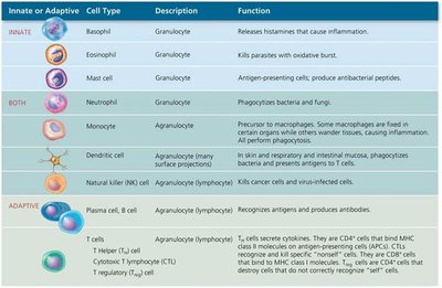
Innate or Adaptive | Cell Type | Description | Function |
|---|---|---|---|
Innate | Basophil | Granulocyte | Releases histamines that cause inflammation. |
Innate | Eosinophil | Granulocyte | Kills parasites with oxidative burst. |
Innate | Mast cell | Granulocyte | Antigen-presenting cells; produce antibacterial peptides. |
Both | Neutrophil | Granulocyte | Phagocytizes bacteria and fungi. |
Both | Monocyte | Agranulocyte | Precursor to macrophages; some fixed in certain organs, others wander tissues. |
Both | Dendritic cell | Agranulocyte | Phagocytizes microbes; initiates adaptive immune responses. |
Both | Natural killer (NK) cell | Agranulocyte (lymphocyte) | Kills cancer cells and virus-infected cells. |
Adaptive | Plasma cell, B cell | Agranulocyte (lymphocyte) | Recognizes antigens and produces antibodies. |
Adaptive | T cells | Agranulocyte (lymphocyte) | Cell-mediated immunity; includes helper, cytotoxic, and regulatory T cells. |
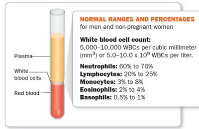
Normal Ranges of White Blood Cells
Neutrophils: 60–70%
Lymphocytes: 20–25%
Monocytes: 3–8%
Eosinophils: 2–4%
Basophils: 0.5–1%
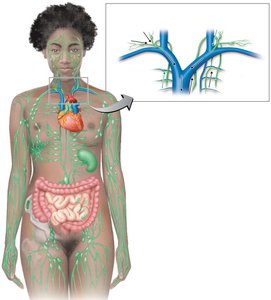
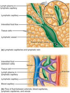
The Lymphoid System
The lymphoid system consists of lymph plasma, lymphatic vessels, lymphoid tissues and organs, and red bone marrow. Lymphoid tissue contains lymphocytes and phagocytic cells. Lymph transports microbes to lymph nodes, where immune cells encounter and destroy pathogens.
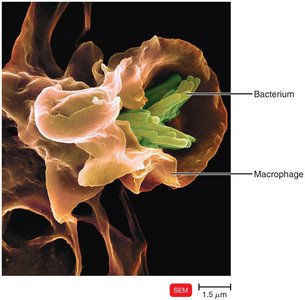
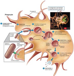
Phagocytosis
Phagocytes and Their Roles
Phagocytes are cells that ingest and destroy microbes and debris. "Professional" phagocytes include neutrophils, eosinophils, and macrophages (derived from monocytes). Macrophages can be fixed (residing in tissues) or free (wandering in tissues).
The Mechanism of Phagocytosis
Chemotaxis: Chemical signals attract phagocytes to the site of infection.
Adherence: Phagocyte attaches to the microbe, often enhanced by opsonization (coating with serum proteins like antibodies or complement).
Ingestion: Pseudopods extend to engulf the microbe, forming a phagosome.
Digestion: Lysosomes fuse with the phagosome to form a phagolysosome, where the microbe is destroyed. Indigestible material is expelled by exocytosis.
Inflammation
Definition and Functions
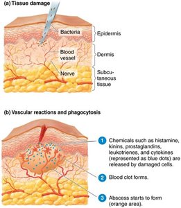
Inflammation is a local defensive response to tissue damage caused by infection, physical, or chemical agents. Its main functions are to destroy or contain the injurious agent and to repair damaged tissue.
Signs and Symptoms (PRISH): Pain, Redness, Immobility, Swelling, Heat.
Types: Acute (rapid, short-term) and chronic (slow, long-term).
Stages of Inflammation
Vasodilation and Increased Permeability: Blood vessels dilate and become more permeable, allowing immune cells and proteins to reach the site of injury. Mediators include histamine, kinins, prostaglandins, and leukotrienes.
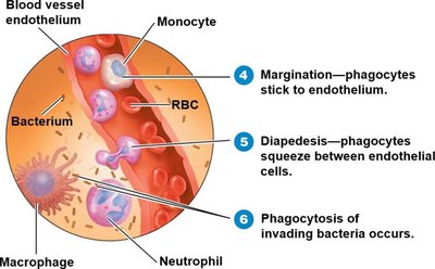
Phagocyte Migration and Phagocytosis: Neutrophils and monocytes migrate to the site, adhere to endothelium (margination), squeeze through vessel walls (diapedesis), and phagocytize microbes.
Tissue Repair: Begins after harmful substances are removed; involves repair of stroma (supporting tissue) and parenchyma (functional tissue).
Vasoactive Mediators of Inflammation
Vasoactive Mediator | Source | Effect |
|---|---|---|
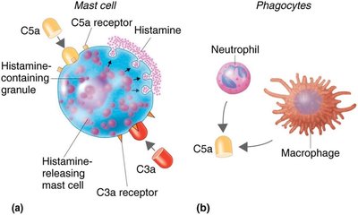
Histamine | Mast cells, basophils, platelets | Vasodilation, increased permeability |
Kinins | Blood plasma | Vasodilation, increased permeability, chemotaxis |
Prostaglandins | Damaged cells | Intensify histamine/kinin effects, help phagocytes move |
Leukotrienes | Mast cells, basophils | Increase permeability, help phagocyte attachment |
Complement | Blood plasma | Stimulates histamine release, attracts phagocytes, promotes phagocytosis |
Cytokines | Macrophages | Vasodilation, increased permeability |
Fever
Mechanism and Effects
Fever is an abnormally high body temperature, usually caused by infection. Cytokines reset the hypothalamic thermostat, leading to increased body temperature. Fever enhances immune function but can be dangerous if too high.
Benefits: Enhances phagocyte and T cell activity, increases production of antimicrobial substances, slows pathogen growth, speeds tissue repair.
Complications: Tachycardia, acidosis, dehydration, seizures, coma, death at extreme temperatures.
Antimicrobial Substances
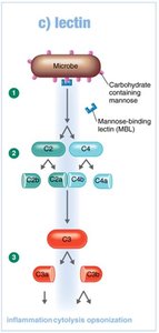
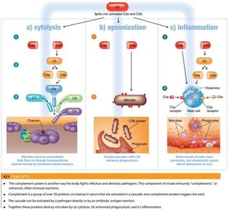
The Complement System
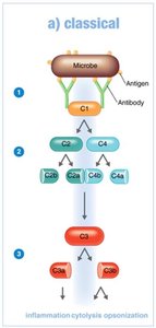
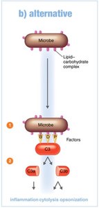
The complement system consists of over 30 serum proteins that enhance immune responses. It can be activated by three pathways: classical (antibody-dependent), alternative (direct binding to microbe), and lectin (mannose-binding lectin). The main outcomes are cytolysis, opsonization, and inflammation.
Cytolysis: Formation of membrane attack complex (MAC) that lyses microbes.
Opsonization: Coating of microbes to enhance phagocytosis.
Inflammation: Complement proteins trigger release of histamine and attract phagocytes.
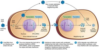
Interferons
Interferons (IFNs) are cytokines with antiviral activity. IFN-α and IFN-β are produced by virus-infected cells and induce neighboring cells to produce antiviral proteins (AVPs) that inhibit viral replication. IFN-γ activates neutrophils and macrophages to kill bacteria.
Iron-Binding Proteins
Iron-binding proteins (transferrin, lactoferrin, ferritin, hemoglobin) sequester iron, limiting its availability to pathogens. Some bacteria produce siderophores to compete for iron.
Antimicrobial Peptides
Antimicrobial peptides are short proteins produced in response to microbial molecules. They have broad-spectrum activity, inhibiting cell wall synthesis, forming pores in membranes, and destroying nucleic acids. Examples include dermcidin, defensins, cathelicidins, and thrombocidin.
Other Factors Affecting Immunity
Genetic Resistance: Certain genetic traits (e.g., sickle cell trait) confer resistance to specific pathogens.
Age: The very young and elderly are more susceptible to infection.
Healthy Practices: Hand hygiene, respiratory etiquette, and safe sex practices reduce disease risk.
