 Back
Back16 Innate Immunity: Nonspecific Defenses of the Host
Study Guide - Smart Notes
 Tailored notes based on your materials, expanded with key definitions, examples, and context.
Chapter 16: Innate Immunity – Nonspecific Defenses of the Host
Introduction to Immunity
Immunity refers to the body's ability to resist or eliminate potentially harmful foreign materials or abnormal cells. The immune system is divided into two main branches: innate immunity (nonspecific, present at birth) and adaptive immunity (specific, acquired after exposure to pathogens). Innate immunity provides the first and second lines of defense, while adaptive immunity provides the third line of defense, characterized by specificity and memory.
Big Picture: Innate vs. Adaptive Immunity
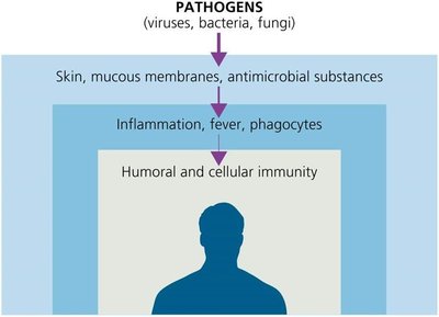
Overview of Immune Defenses
First Line of Defense: Skin, mucous membranes, and antimicrobial substances prevent pathogen entry.
Second Line of Defense: Inflammation, fever, and phagocytes act rapidly and nonspecifically.
Third Line of Defense: Humoral and cellular immunity (adaptive), slower but highly specific and has memory.
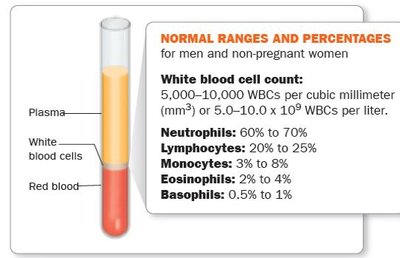
Blood Cell Counts and Health
White blood cell (WBC) counts are important diagnostic tools. High WBC counts may indicate bacterial infections or autoimmune diseases, while low counts may suggest viral infections or other conditions. The normal range and distribution of WBCs are as follows:
The Concept of Immunity
Definitions and Types
Immunity (Resistance): The ability to ward off disease.
Susceptibility: Lack of resistance to a disease.
Innate Immunity: Present at birth, rapid, nonspecific, no memory.
Adaptive Immunity: Specific, slower to respond, has memory, involves lymphocytes (T and B cells).
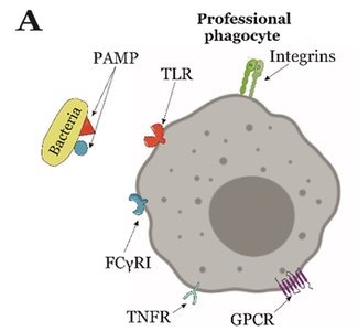
Toll-like Receptors (TLRs) and Pathogen Recognition
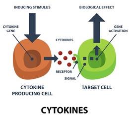
Innate immune responses are triggered by protein receptors in the plasma membrane of defensive cells. Toll-like receptors (TLRs) on host cells recognize pathogen-associated molecular patterns (PAMPs) such as lipopolysaccharide (LPS), flagellin, and peptidoglycan. Binding of TLRs to PAMPs induces the release of cytokines, which regulate immune responses.
First Line of Defense: Skin and Mucous Membranes
Physical Factors
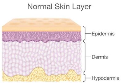
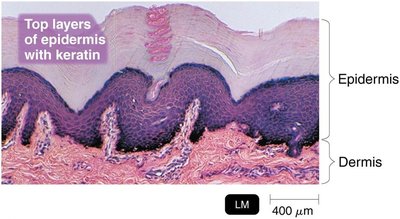
The skin and mucous membranes act as physical barriers to pathogen entry. The skin consists of the dermis (connective tissue) and the epidermis (tightly packed epithelial cells with keratin). Shedding and dryness of the skin inhibit microbial growth.
Mucous Membranes: Line the gastrointestinal, respiratory, and genitourinary tracts. Mucus traps microbes and prevents drying.
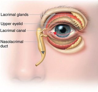
Lacrimal Apparatus: Produces tears that wash the eye and drain into the nasolacrimal duct.
Ciliary Escalator: Cilia in the respiratory tract move mucus and trapped microbes away from the lungs.
Other Mechanisms: Earwax, urine flow, vaginal secretions, peristalsis, defecation, vomiting, and diarrhea help remove microbes.
Chemical Factors
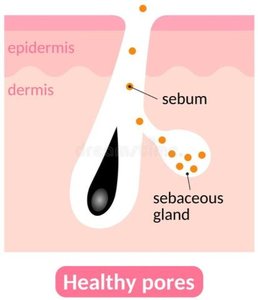
Sebum: Forms a protective film and lowers skin pH (3–5), inhibiting microbial growth.
Lysozyme: Enzyme in sweat, tears, saliva, and urine that destroys bacterial cell walls.
Low pH: Gastric juice (pH 1.2–3.0) and vaginal secretions (pH 3–5) inhibit or destroy microbes.
Normal Microbiota and Innate Immunity
Microbial Antagonism (Competitive Exclusion): Normal microbiota compete with pathogens for space and nutrients, produce substances harmful to pathogens, and alter environmental conditions.
Commensalism: One organism benefits, the other is unharmed.
Probiotics: Live microbial cultures that confer health benefits, such as lactic acid bacteria preventing diarrhea and pathogen colonization.

Second Line of Defense: Cellular Components
Formed Elements in Blood
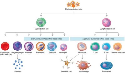
Blood consists of plasma and formed elements (cells and cell fragments). The main formed elements are erythrocytes (RBCs), leukocytes (WBCs), and platelets. These are produced in red bone marrow via hematopoiesis.

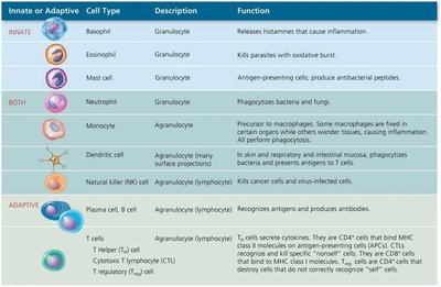
Leukocytes: Granulocytes and Agranulocytes
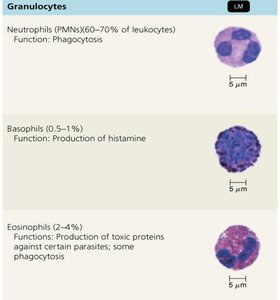
Granulocytes: Visible granules in cytoplasm. Types include:
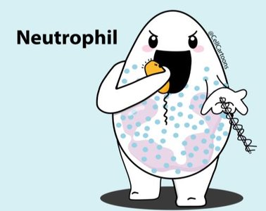
Neutrophils: Phagocytic, early infection responders.
Basophils: Release histamine, involved in allergic responses.
Eosinophils: Phagocytic, toxic against parasites and helminths.
Agranulocytes: No visible granules. Types include:
Monocytes: Mature into macrophages, phagocytic.
Dendritic Cells: Phagocytic, initiate adaptive immunity.
Lymphocytes: T cells, B cells, and NK cells; involved in adaptive immunity.
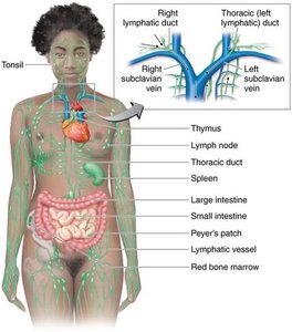
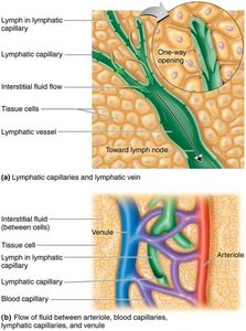
The Lymphatic System
Structure and Function
The lymphatic system consists of lymph, lymphatic vessels, lymphoid tissue, and red bone marrow. It contains lymphocytes and phagocytic cells. Lymph carries microbes to lymph nodes, where they are destroyed by lymphocytes and macrophages.
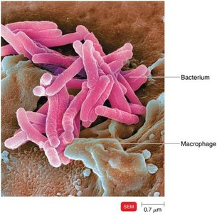
Phagocytes and Phagocytosis
Definition and Types
Phagocytes: Cells that ingest and destroy microbes and debris. Main types are neutrophils and macrophages.
Fixed Macrophages: Reside in specific tissues and organs.
Free (Wandering) Macrophages: Move throughout tissues and gather at infection sites.
Mechanism of Phagocytosis
Chemotaxis: Chemical attraction of phagocytes to microorganisms.
Adherence: Attachment of phagocyte to microbe surface.
Ingestion: Microbe is engulfed; opsonization (coating with serum proteins) enhances ingestion.
Digestion: Microbe is digested inside a phagolysosome.
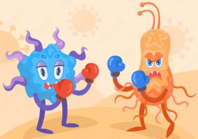
Inflammation
Purpose and Signs
Inflammation is a local defensive response to tissue damage. Its main functions are to destroy the injurious agent, limit its effects, and repair damaged tissue. Signs include pain, redness, immobility, swelling (edema), and heat.
Stages and Mediators
Vasodilation: Increases blood flow to the area.
Increased Permeability: Allows defensive substances to leave the blood and enter tissues.
Chemical Mediators: Histamine, kinins, prostaglandins, leukotrienes, and cytokines.
Vasoactive Mediator | Source | Effect |
|---|---|---|
Histamine | Mast cells, basophils, platelets | Vasodilation, increased permeability |
Kinins | Blood plasma | Chemotaxis, attract neutrophils |
Prostaglandins | Damaged cells | Intensify histamine/kinin effects, help phagocytes move |
Leukotrienes | Mast cells, basophils | Increase permeability, help phagocyte attachment |
Complement | Blood plasma | Stimulates histamine release, attracts phagocytes, promotes phagocytosis |
Cytokines | Fixed macrophages | Vasodilation, increased permeability |
Phagocyte Migration and Tissue Repair
Margination: Phagocytes stick to blood vessel walls in response to cytokines.
Diapedesis: Phagocytes squeeze between endothelial cells to reach damaged tissue.
Tissue Repair: Final stage; stroma (supporting tissue) and parenchyma (functioning tissue) produce new cells.
Fever
Cause and Effects
Fever is an abnormally high body temperature, usually caused by infection. Cytokines cause the hypothalamus to reset to a higher temperature, which is maintained until the cytokines are eliminated. Fever enhances immune responses and inhibits some pathogens.
Antimicrobial Substances
The Complement System
The complement system consists of over 30 proteins produced by the liver, acting in a cascade to enhance immune responses. The goal is activation of C3, leading to cytolysis, opsonization, and inflammation.
Classical Pathway: Triggered by antibodies binding to antigens.
Alternative Pathway: Triggered by complement proteins binding directly to pathogen surfaces.
Lectin Pathway: Triggered by lectin binding to microbial carbohydrates.
Outcome | Description |
|---|---|
Cytolysis | Membrane attack complex (MAC) forms, lysing microbe |
Opsonization | Coating of microbe to enhance phagocytosis |
Inflammation | Complement proteins trigger histamine release |
Interferons
IFN-α and IFN-β: Produced in response to viral infections; induce antiviral proteins in neighboring cells.
IFN-γ: Activates neutrophils and macrophages to kill bacteria.
Iron-Binding Proteins
Transport and store iron, depriving pathogens of this essential nutrient.
Examples: transferrin, lactoferrin, ferritin, hemoglobin.
Pathogens may produce siderophores to compete for iron.
Antimicrobial Peptides (AMPs)
Short peptides produced in response to microbial molecules.
Broad spectrum: inhibit cell wall synthesis, form pores, destroy DNA/RNA.
Other Factors Affecting Immunity
Genetic Resistance: Certain genetic traits confer resistance (e.g., sickle cell trait and malaria).
Age: Very young and elderly are more susceptible to infection.
Hygiene: Practices like handwashing reduce infection risk.
