 Back
BackInnate Immunity: Nonspecific Defenses of the Host
Study Guide - Smart Notes
 Tailored notes based on your materials, expanded with key definitions, examples, and context.
Innate Immunity: Nonspecific Defenses of the Host
Overview of Immunity
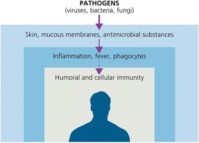
Immunity refers to the body's ability to resist or eliminate potentially harmful foreign materials or abnormal cells. The immune system is divided into two main branches: innate immunity (nonspecific, present at birth, rapid response) and adaptive immunity (specific, slower, memory component). Innate immunity provides the first and second lines of defense against pathogens, while adaptive immunity provides the third line of defense.
Innate Immunity: Rapid, nonspecific, no memory. Includes barriers, phagocytes, inflammation, and fever.
Adaptive Immunity: Slower, specific, has memory. Involves lymphocytes (T cells and B cells).
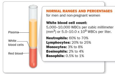
Blood Cell Counts and Health
White blood cell (WBC) counts are important diagnostic tools. Abnormal counts can indicate infections, immune disorders, or other health conditions.
High WBC count: May indicate bacterial infection, autoimmune disease, or medication side effects.
Low WBC count: May indicate viral infection, pneumonia, autoimmune disease, or cancer.
The Concept of Immunity
Definitions and Types
Immunity (Resistance): Ability to ward off disease.
Susceptibility: Lack of resistance to disease.
Innate Immunity: Present at birth, rapid, nonspecific, no memory.
Adaptive Immunity: Specific, slower, memory component, involves T and B lymphocytes.
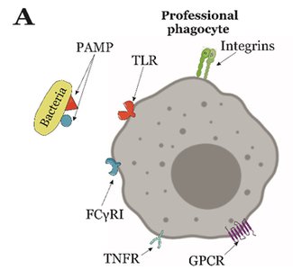
Toll-like Receptors (TLRs) and Pathogen Recognition
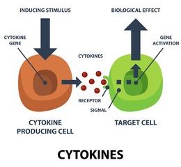
Innate immune responses are triggered by protein receptors in the plasma membrane of defensive cells. Toll-like receptors (TLRs) on host cells recognize pathogen-associated molecular patterns (PAMPs) such as lipopolysaccharide (LPS), flagellin, and peptidoglycan. Binding of TLRs to PAMPs induces the release of cytokines, which regulate immune responses.
First Line of Defense: Skin and Mucous Membranes
Physical Factors
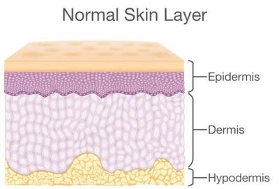
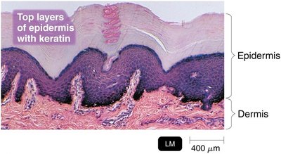
The skin and mucous membranes act as physical barriers to prevent pathogen entry. The skin consists of the epidermis (outer layer, keratinized, tightly packed cells) and the dermis (inner connective tissue layer). Shedding and dryness of skin inhibit microbial growth.
Skin: Largest organ, physical barrier, keratinized epidermis.
Mucous membranes: Line gastrointestinal, respiratory, and genitourinary tracts; secrete mucus to trap microbes.
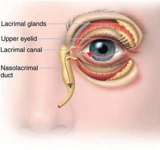
Lacrimal apparatus: Produces tears to wash microbes from the eye.
Additional Physical Defenses
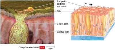
Ciliary escalator: Cilia in the respiratory tract move mucus and trapped microbes away from the lungs.
Earwax: Prevents entry of microbes and debris.
Urine and vaginal secretions: Flush out microbes.
Peristalsis, defecation, vomiting, diarrhea: Expel pathogens from the gastrointestinal tract.
Chemical Factors
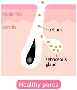
Sebum: Oily substance from sebaceous glands; forms a protective film and lowers skin pH (3-5).
Lysozyme: Enzyme in sweat, tears, saliva, and urine; destroys bacterial cell walls (peptidoglycan).
Low pH: Gastric juice (pH 1.2-3.0) and vaginal secretions (pH 3-5) inhibit microbial growth.
Normal Microbiota and Innate Immunity
Microbial antagonism (competitive exclusion): Normal microbiota compete with pathogens for space and nutrients, produce substances harmful to pathogens, and alter environmental conditions.
Commensalism: One organism benefits, the other is unharmed.
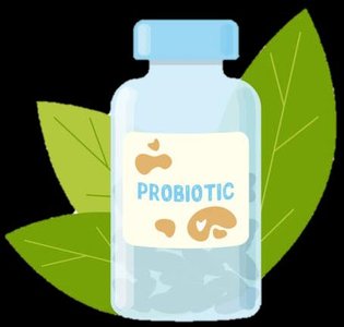
Probiotics: Live microbial cultures that confer health benefits, such as lactic acid bacteria preventing diarrhea and pathogen colonization.
Second Line of Defense: Cellular Components
Formed Elements in Blood
Blood consists of plasma and formed elements (cells and cell fragments). The main formed elements are erythrocytes (red blood cells), leukocytes (white blood cells), and platelets. All blood cells originate from stem cells in red bone marrow through hematopoiesis.

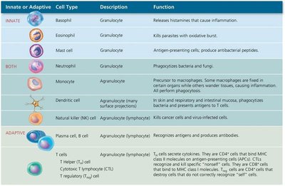
Leukocytes: Types and Functions
Leukocytes are divided into granulocytes (with visible granules) and agranulocytes (without visible granules).
Innate or Adaptive | Cell Type | Description | Function |
|---|---|---|---|
Innate | Basophil | Granulocyte | Releases histamines that cause inflammation |
Innate | Eosinophil | Granulocyte | Kills parasites with oxidative burst |
Innate | Mast cell | Granulocyte | Antigen-presenting cell; produces antibacterial peptides |
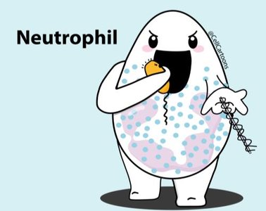
Both | Neutrophil | Granulocyte | Phagocytizes bacteria and fungi |
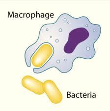
Both | Monocyte | Agranulocyte | Precursor to macrophages; phagocytic |
Both | Dendritic cell | Agranulocyte | Phagocytosis and initiation of adaptive immune responses |
Both | Natural killer (NK) cell | Agranulocyte | Kills cancer cells and virus-infected cells |
Adaptive | Plasma cell, B cell | Agranulocyte | Produces antibodies |
Adaptive | T cells | Agranulocyte | Cell-mediated immunity |
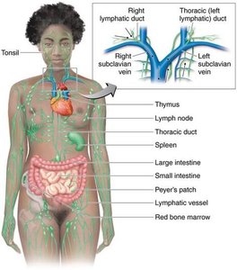
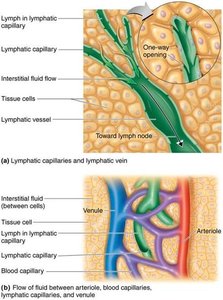
The Lymphatic System
The lymphatic system consists of lymph, lymphatic vessels, lymphoid tissue, and red bone marrow. It contains lymphocytes and phagocytic cells. Lymph carries microbes to lymph nodes, where they are destroyed by lymphocytes and macrophages.
Phagocytes and Phagocytosis
Phagocytes
Phagocytes are cells that ingest and destroy microbes and debris. Types include neutrophils, macrophages (mature monocytes), and dendritic cells. Fixed macrophages reside in tissues, while free (wandering) macrophages move to sites of infection.
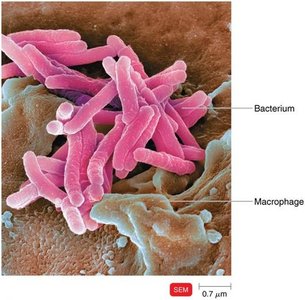
Mechanism of Phagocytosis
Phagocytosis occurs in four main phases:
Chemotaxis: Phagocytes are attracted to microbes by chemical signals.
Adherence: Phagocyte attaches to the microbe's surface.
Ingestion: Microbe is engulfed into a phagosome; opsonization (coating with serum proteins) enhances ingestion.
Digestion: Microbe is destroyed inside a phagolysosome.
Inflammation
Stages and Functions
Inflammation is a local defensive response to tissue damage. The main signs are pain, redness, immobility, swelling (edema), and heat (PRISH). Functions include destroying the injurious agent, limiting its effects, and repairing tissue.
Vasodilation: Increases blood flow to the area (redness, heat).
Increased permeability: Allows defensive substances to enter tissues (swelling, pain).
Chemotaxis: Attracts phagocytes to the site of injury.
Vasoactive Mediator | Source | Effect |
|---|---|---|
Histamine | Mast cells, basophils, platelets | Vasodilation, increased permeability |
Kinins | Blood plasma | Chemotaxis, attract neutrophils |
Prostaglandins | Damaged cells | Intensify histamine/kinin effects, help phagocytes move through capillary walls |
Leukotrienes | Mast cells, basophils | Increase permeability, help phagocytes attach to pathogens |
Complement | Blood plasma | Stimulates histamine release, attracts phagocytes, promotes phagocytosis |
Cytokines | Fixed macrophages | Vasodilation, increased permeability |
Phagocyte Migration and Tissue Repair
Margination: Phagocytes stick to blood vessel walls at the site of inflammation.
Diapedesis: Phagocytes squeeze through endothelial cells to reach damaged tissue.
Tissue repair: Final stage; stroma (supporting tissue) and parenchyma (functioning tissue) produce new cells.
Fever
Causes and Effects
Fever is an abnormally high body temperature, usually caused by infection. Cytokines trigger the hypothalamus to raise body temperature. Fever enhances immune responses and inhibits some pathogens.
Benefits: Increases production of immune cells, inhibits growth of some microbes, speeds up tissue repair.
Antimicrobial Substances
The Complement System
The complement system is a group of over 30 proteins produced by the liver that enhance immune responses. Complement activation occurs via three pathways:
Classical pathway: Triggered by antibodies binding to antigens.
Alternative pathway: Triggered by direct interaction with pathogen surfaces.
Lectin pathway: Triggered by lectin binding to microbial carbohydrates.
Outcomes: Cytolysis (cell lysis), opsonization (enhanced phagocytosis), inflammation.
Regulation: Regulatory proteins prevent host cell damage.
Deficiencies: Increase susceptibility to infections.
Pathogen evasion: Capsules can prevent complement activation.
Interferons
Interferons (IFNs) are cytokines with antiviral activity. Types include:
IFN-α and IFN-β: Produced in response to viral infection; induce antiviral proteins in neighboring cells.
IFN-γ: Activates neutrophils and macrophages to kill bacteria.
Iron-Binding Proteins
Iron-binding proteins (e.g., transferrin, lactoferrin, ferritin, hemoglobin) sequester iron, limiting its availability to pathogens. Some bacteria produce siderophores to compete for iron.
Antimicrobial Peptides (AMPs)
AMPs are short peptides produced in response to microbial molecules. They have broad-spectrum activity, inhibiting cell wall synthesis, forming pores in membranes, and destroying DNA/RNA.
Other Factors Affecting Immunity
Genetic resistance: Certain genetic traits (e.g., sickle cell trait) confer resistance to specific pathogens.
Age: Very young and elderly individuals are more susceptible to infection.
Hygiene: Practices like handwashing reduce infection risk.
