 Back
BackInnate Immunity: Nonspecific Defenses of the Host
Study Guide - Smart Notes
 Tailored notes based on your materials, expanded with key definitions, examples, and context.
Innate Immunity: Nonspecific Defenses of the Host
Overview of Immunity
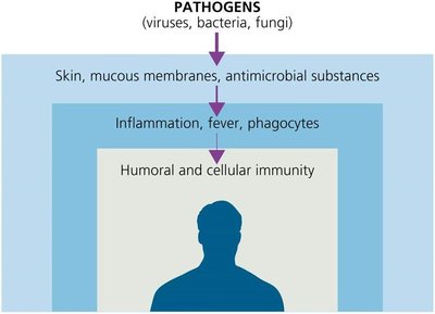
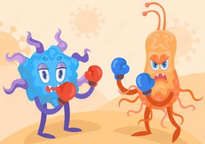
Immunity refers to the body's ability to resist or eliminate potentially harmful foreign materials or abnormal cells. The immune system is divided into two main branches: innate immunity (nonspecific, present at birth, rapid response) and adaptive immunity (specific, slower, memory component). Innate immunity provides the first and second lines of defense against pathogens, while adaptive immunity provides the third line of defense.
Key Differences: Innate vs. Adaptive Immunity
Innate Immunity: Rapid, nonspecific, no memory, includes barriers (skin, mucous membranes), phagocytes, inflammation, and fever.
Adaptive Immunity: Slower, specific to pathogens, has memory, involves lymphocytes (T cells and B cells).
First Line of Defense: Physical and Chemical Barriers
Physical Barriers
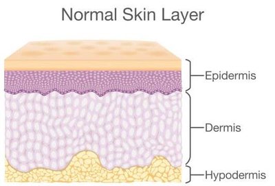
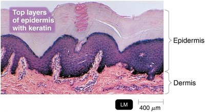
The skin and mucous membranes act as the body's primary physical barriers to infection. These structures prevent the entry of pathogens through tightly packed epithelial cells and protective secretions.
Skin: Composed of the outer epidermis (keratinized, tightly packed cells) and inner dermis (connective tissue). Shedding and dryness inhibit microbial growth.
Mucous Membranes: Line the gastrointestinal, respiratory, and genitourinary tracts. Secrete mucus to trap microbes and prevent drying.
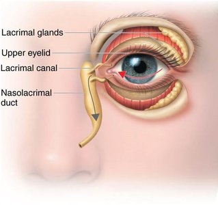
Lacrimal Apparatus: Produces tears that wash microbes from the eye surface.
Ciliary Escalator: Cilia in the respiratory tract move mucus and trapped particles away from the lungs.
Other Physical Factors: Earwax, urine flow, vaginal secretions, peristalsis, defecation, vomiting, and diarrhea help remove microbes from the body.
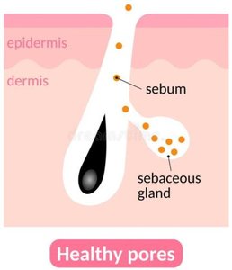
Chemical Barriers
Chemical factors enhance the effectiveness of physical barriers by creating hostile environments for pathogens.
Sebum: Oily substance produced by sebaceous glands; forms a protective film and lowers skin pH (3–5).
Lysozyme: Enzyme in sweat, tears, saliva, and urine that destroys bacterial cell walls (peptidoglycan).
Low pH: Gastric juice (pH 1.2–3.0) and vaginal secretions (pH 3–5) inhibit microbial growth.
Role of Normal Microbiota in Innate Immunity
Normal microbiota protect the host by competing with pathogens for nutrients and space (microbial antagonism), producing substances harmful to pathogens, and altering environmental conditions. Some microbes are commensal (benefit without harming the host), while probiotics are live cultures administered for health benefits.
Second Line of Defense: Cellular Components and Processes
Formed Elements in Blood
Blood contains plasma and formed elements: erythrocytes (red blood cells), leukocytes (white blood cells), and platelets. Leukocytes are crucial for immune defense and are produced in red bone marrow via hematopoiesis.

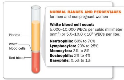
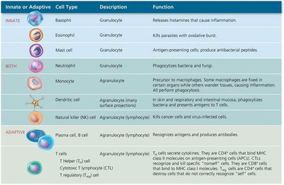
Types of Leukocytes
Leukocytes are classified as granulocytes or agranulocytes based on the presence of cytoplasmic granules.
Innate or Adaptive | Cell Type | Description | Function |
|---|---|---|---|
Innate | Basophil | Granulocyte | Releases histamines that cause inflammation |
Innate | Eosinophil | Granulocyte | Kills parasites with oxidative burst |
Innate | Mast cell | Granulocyte | Antigen-presenting cells; produce antibacterial peptides |
Both | Neutrophil | Granulocyte | Phagocytizes bacteria and fungi |
Both | Monocyte | Agranulocyte | Precursor to macrophages; some fixed in certain organs, others wander tissues |
Both | Dendritic cell | Agranulocyte | In skin and mucosa; phagocytizes bacteria and viruses |
Both | Natural killer (NK) cell | Agranulocyte | Kills cancer cells and virus-infected cells |
Adaptive | Plasma cell, B cell | Agranulocyte | Recognizes antigens and produces antibodies |
Adaptive | T cells | Agranulocyte | Cell-mediated immunity |
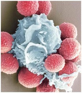
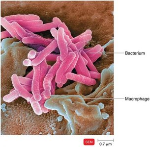
Phagocytes and Phagocytosis
Phagocytes are cells that ingest and destroy microbes and debris. Major phagocytes include neutrophils and macrophages (derived from monocytes). Phagocytosis involves four main steps: chemotaxis, adherence, ingestion (often enhanced by opsonization), and digestion within phagolysosomes.
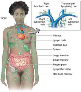
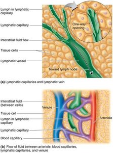
The Lymphatic System
The lymphatic system consists of lymph, lymphatic vessels, lymphoid tissue, and red bone marrow. It transports lymph (containing microbes) to lymph nodes, where lymphocytes and macrophages destroy pathogens. The system is essential for immune surveillance and fluid balance.
Inflammation
Inflammation is a local defensive response to tissue damage, characterized by pain, redness, immobility, swelling, and heat (PRISH). Its functions are to destroy or contain injurious agents and repair damaged tissue. Key mediators include histamine, kinins, prostaglandins, leukotrienes, and cytokines, which promote vasodilation and increased vascular permeability.
Vasoactive Mediator | Source | Effect |
|---|---|---|
Histamine | Mast cells, basophils, platelets | Vasodilation, increased permeability |
Kinins | Blood plasma | Chemotaxis, attracts neutrophils |
Prostaglandins | Damaged cells | Intensify histamine/kinin effects, help phagocytes move through capillary walls |
Leukotrienes | Mast cells, basophils | Increase permeability, help phagocyte attachment |
Complement | Blood plasma | Stimulates histamine release, attracts phagocytes, promotes phagocytosis |
Cytokines | Fixed macrophages | Vasodilation, increased permeability |
Fever
Fever is an abnormally high body temperature, usually caused by infection. Cytokines trigger the hypothalamus to raise body temperature, which can inhibit pathogen growth, increase immune reactions, and speed up tissue repair. When the fever 'breaks,' vasodilation and sweating return the body to normal temperature.
Antimicrobial Substances
The Complement System
The complement system is a group of over 30 proteins that enhance immune responses. Complement activation occurs via three pathways: classical (antibody-dependent), alternative (direct pathogen contact), and lectin (mannose-binding). The main outcomes are cytolysis (cell lysis), opsonization (enhanced phagocytosis), and inflammation.
Classical Pathway: Triggered by antibodies binding to antigens.
Alternative Pathway: Triggered by complement proteins binding directly to pathogen surfaces.
Lectin Pathway: Triggered by lectin binding to microbial carbohydrates.
Interferons
Interferons (IFNs) are cytokines with antiviral activity. IFN-α and IFN-β are produced in response to viral infections and induce neighboring cells to produce antiviral proteins. IFN-γ activates neutrophils and macrophages to kill bacteria.
Iron-Binding Proteins
Iron-binding proteins (e.g., transferrin, lactoferrin, ferritin, hemoglobin) sequester iron, limiting its availability to pathogens. Some bacteria produce siderophores to compete for iron.
Antimicrobial Peptides (AMPs)
AMPs are short peptides produced in response to microbial molecules. They have broad-spectrum activity, including inhibition of cell wall synthesis, pore formation, and destruction of microbial DNA/RNA.
Other Factors Affecting Resistance
Genetic resistance: Certain genetic traits (e.g., sickle cell trait) can confer resistance to specific pathogens.
Age: The very young and elderly are more susceptible to infection.
Hygiene: Practices such as handwashing reduce infection risk.
