 Back
Innate Immunity: Nonspecific Defenses of the Host
Study Guide - Smart Notes
 Tailored notes based on your materials, expanded with key definitions, examples, and context.
Innate Immune Response
Overview of Innate Defenses
The innate immune system provides the body's first and immediate line of defense against invading pathogens. It consists of physical barriers, sensor systems, cellular defenses, and chemical mediators that act rapidly and nonspecifically to prevent infection and eliminate invaders.
First line of defense: Physical and chemical barriers that prevent pathogen entry.
Sensor systems: Cellular receptors and proteins that detect breaches in barriers and initiate immune responses.
Effector actions: Cellular and molecular mechanisms that destroy or remove invaders.
First Line of Defense
Physical Barriers
Physical barriers are anatomical structures that separate the internal environment from the external world, preventing pathogen entry.
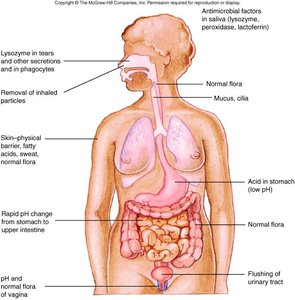
Skin: The most visible and effective barrier, composed of the tough dermis and keratinized epidermis. The outer layers slough off, removing attached microbes.
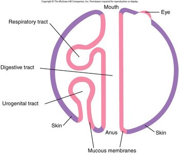
Mucous membranes: Line the digestive, respiratory, and genitourinary tracts. Mucus traps microbes, and cilia or other mechanisms help expel them.
Chemical Barriers and Antimicrobial Substances
Both skin and mucous membranes are protected by a variety of antimicrobial substances:
Lysozyme: Enzyme that degrades peptidoglycan; found in tears, saliva, and phagocytes.
Peroxidase: Breaks down hydrogen peroxide to produce reactive oxygen; found in saliva, tissues, and phagocytes.
Lactoferrin: Sequesters iron, inhibiting microbial growth; present in saliva, phagocytes, blood, and tissue fluids.
Defensins: Antimicrobial peptides that disrupt microbial membranes; found on mucous membranes and in phagocytes.
Normal Flora
Normal flora are non-pathogenic microorganisms that colonize body surfaces and provide protection by:
Competing with pathogens for binding sites and nutrients (competitive exclusion).
Producing substances that inhibit or kill pathogens.
Additional info: Disruption of normal flora (e.g., by antibiotics) can increase susceptibility to infections.
Sensor Systems
Pattern Recognition Receptors (PRRs)
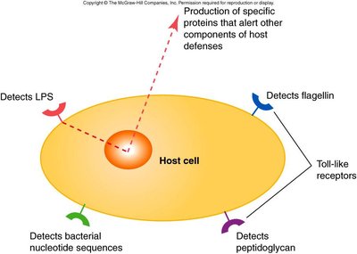
Sensor systems detect signs of tissue damage or microbial invasion using pattern recognition receptors (PRRs), which recognize conserved microbial molecules.
Toll-like receptors (TLRs): Located on cell surfaces or endosomes; detect microbial components such as lipopolysaccharide (LPS), flagellin, peptidoglycan, and bacterial nucleic acids.
NOD proteins: Detect microbial molecules inside the cell.
The Complement System
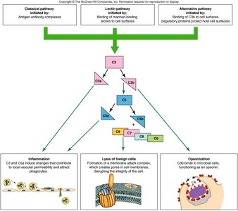
The complement system is a group of proteins circulating in blood and tissue fluids in an inactive form. Activation triggers a cascade of reactions leading to pathogen destruction. There are three main pathways of activation:
Alternative pathway: Triggered by C3b binding to microbial surfaces.
Lectin pathway: Initiated by mannose-binding lectins (MBL) binding to microbial carbohydrates.
Classical pathway: Initiated by antibodies binding to antigens.
Major Outcomes of Complement Activation
Inflammation: C3a and C5a induce vascular changes and attract immune cells.
Opsonization: C3b coats microbes, enhancing phagocytosis.
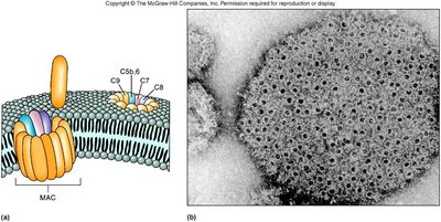
Lysis of foreign cells: Formation of the membrane attack complex (MAC) creates pores in microbial membranes, leading to cell lysis (especially effective against Gram-negative bacteria).
Cells of the Immune System
Blood Cell Formation (Hematopoiesis)
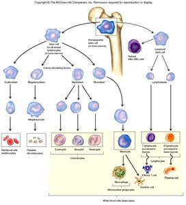
All blood cells, including immune cells, originate from hematopoietic stem cells in the bone marrow. Differentiation is regulated by colony-stimulating factors.
Red blood cells (erythrocytes): Carry oxygen.
Platelets: Involved in blood clotting.
White blood cells (leukocytes): Key players in host defense; divided into granulocytes, mononuclear phagocytes, dendritic cells, and lymphocytes.
Granulocytes
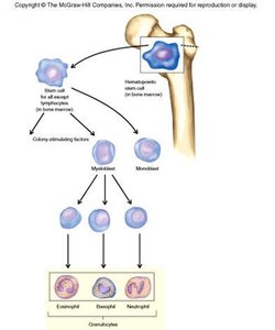
Granulocytes are white blood cells with cytoplasmic granules. They include:
Neutrophils: Most abundant; primary phagocytes in innate immunity.
Basophils: Involved in allergic responses.
Eosinophils: Important in defense against parasitic worms and in allergic reactions.
Mononuclear Phagocytes and Dendritic Cells
Monocytes: Circulate in blood; differentiate into macrophages in tissues.
Macrophages: Abundant in tissues such as liver, spleen, lymph nodes, and lungs; phagocytose microbes and debris.
Dendritic cells: Function as antigen-presenting cells, bridging innate and adaptive immunity by capturing antigens and presenting them to lymphocytes.
Lymphocytes
B lymphocytes (B cells): Produce antibodies; central to adaptive immunity.
T lymphocytes (T cells): Mediate cellular immunity.
Natural killer (NK) cells: Destroy infected or abnormal host cells without antigen specificity.
Cell Communication in Immunity
Surface Receptors
Surface receptors are membrane proteins that bind specific signaling molecules (ligands), transmitting signals into the cell to initiate responses.
Cytokines
Cytokines are chemical messengers that regulate immune cell function. Major classes include:
Chemokines: Direct cell movement (chemotaxis).
Colony-stimulating factors: Stimulate blood cell production and differentiation.
Interferons: Control viral infections and modulate immune responses.
Interleukins: Produced by leukocytes; regulate both innate and adaptive immunity.
Tumor necrosis factor: Induces inflammation and kills tumor cells.
Adhesion Molecules
Adhesion molecules enable immune cells to adhere to each other and to blood vessel walls, facilitating migration to sites of infection or injury.
Phagocytosis
Steps of Phagocytosis
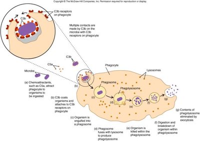
Phagocytosis is the process by which phagocytes engulf and destroy microbes and debris. The steps include:
Chemotaxis: Phagocytes are attracted to infection sites by chemical signals.
Recognition and attachment: Phagocytes bind to microbes using receptors (often enhanced by opsonization with C3b).
Engulfment: The microbe is engulfed into a phagosome.
Phagosome-lysosome fusion: The phagosome fuses with a lysosome, forming a phagolysosome.
Destruction and digestion: Microbes are killed by toxic oxygen products and enzymes.
Exocytosis: Debris is expelled from the cell.
Inflammation
Initiation and Process
Inflammation is a localized response to tissue damage or infection, characterized by heat, pain, redness, swelling, and sometimes loss of function. It is initiated by:
Microbial products triggering TLRs on macrophages, leading to cytokine release.
Complement activation (C3a, C5a) attracting immune cells.
Tissue damage activating enzymatic cascades.
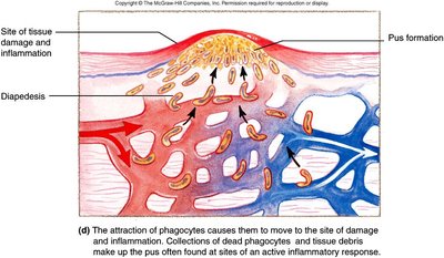
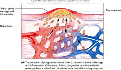
The inflammatory process involves vasodilation, increased vascular permeability, and migration of leukocytes (diapedesis) to the site of injury.
Outcomes of Inflammation
Limits damage and restores tissue function.
Excessive inflammation can cause tissue damage, especially in sensitive organs (e.g., brain, spinal cord).
Apoptosis
Apoptosis is programmed cell death that eliminates cells without triggering inflammation. Apoptotic cells are engulfed by macrophages without inducing an inflammatory response.
Fever
Fever is a systemic response to infection, especially bacterial. It is triggered by pyrogens (endogenous cytokines or exogenous microbial products) and results in:
Inhibition of pathogen growth by raising body temperature above optimal levels for microbes.
Acceleration of immune responses and tissue repair.
