 Back
Innate Immunity: Nonspecific Defenses of the Host – Study Notes
Study Guide - Smart Notes
 Tailored notes based on your materials, expanded with key definitions, examples, and context.
Innate Immunity: Nonspecific Defenses of the Host
Overview of Immunity
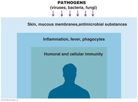
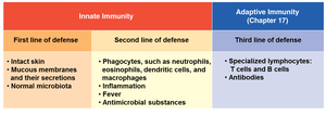
Immunity refers to the body's ability to ward off disease, while susceptibility is the lack of resistance to disease. The immune system is divided into innate immunity (nonspecific, present at birth, rapid response) and adaptive immunity (specific, slower response, memory component). Innate immunity provides the first and second lines of defense against pathogens, including physical barriers, cellular responses, and antimicrobial substances.
The Concept of Immunity
Innate Immunity: Defenses against any pathogen; rapid and present at birth.
Adaptive Immunity: Immunity or resistance to a specific pathogen; slower to respond, has memory component.
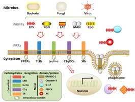
Toll-like receptors (TLRs): Proteins on host cells that recognize pathogen-associated molecular patterns (PAMPs) and trigger cytokine release, regulating immune responses.
White Blood Cell Counts and Types
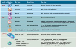
White blood cell (WBC) counts are used to measure leukocytes in the blood. Normal ranges are 5,000–10,000 WBCs per mm3. Different types of WBCs play distinct roles in immunity:
Neutrophils: 60–70%
Lymphocytes: 20–25%
Monocytes: 3–8%
Eosinophils: 2–4%
Basophils: 0.5–1%
First Line of Defense: Skin and Mucous Membranes
Physical Factors
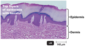
The skin and mucous membranes act as physical barriers to prevent pathogen entry. The skin consists of the dermis (connective tissue) and epidermis (tightly packed epithelial cells containing keratin). Shedding and dryness of skin inhibit microbial growth.
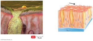
Mucous membranes: Line all body cavities that open to the exterior (respiratory, digestive, urinary, reproductive tracts).
Mucus: Viscous glycoproteins that trap microbes and prevent tracts from drying out.
Ciliary escalator: Transports microbes trapped in mucus away from the lungs.
Other mechanisms: Earwax, urine flow, vaginal secretions, peristalsis, defecation, vomiting, diarrhea.
Chemical Factors
Sebum: Forms a protective film and lowers skin pH (3–5).
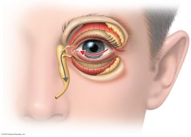
Lysozyme: Enzyme in perspiration, tears, saliva, and urine that destroys bacterial cell walls.
Gastric juice: Low pH (1.2–3.0) destroys most bacteria and toxins.
Vaginal secretions: Low pH (3–5) inhibits microbes.
Role of Normal Microbiota
Microbial antagonism: Normal microbiota compete with pathogens, produce substances harmful to pathogens, and alter conditions affecting pathogen survival.
Commensalism: One organism benefits, the host is unharmed.
Probiotics: Live microbial cultures administered for beneficial effects.
Second Line of Defense: Cellular and Chemical Responses
Formed Elements in Blood
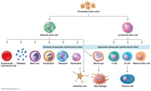
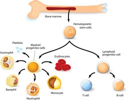
Blood contains cells and cell fragments suspended in plasma, including erythrocytes (red blood cells), leukocytes (white blood cells), and platelets. These are produced in red bone marrow via hematopoiesis.
The Lymphatic System
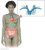
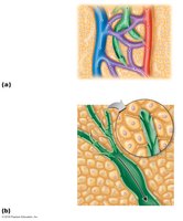
The lymphatic system consists of lymph, lymphatic vessels, lymphoid tissue, and red bone marrow. It contains lymphocytes and phagocytic cells. Lymph carries microbes to lymph nodes, where lymphocytes and macrophages destroy pathogens.
Phagocytes and Phagocytosis
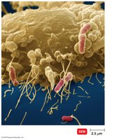
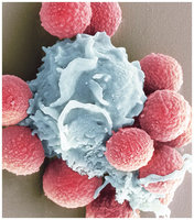
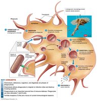
Phagocytes are cells that ingest and destroy microbes. Fixed macrophages reside in tissues and organs, while free macrophages roam tissues and gather at infection sites. The process of phagocytosis includes chemotaxis, adherence, ingestion (opsonization), and digestion.
Inflammation
Stages and Mediators
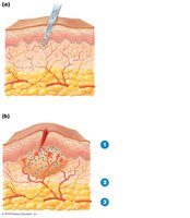
Inflammation is characterized by redness, swelling, pain, and heat. It destroys injurious agents, limits their effects, and repairs damaged tissue. Acute-phase proteins, histamine, kinins, prostaglandins, leukotrienes, and cytokines mediate inflammation.
Phagocyte Migration and Phagocytosis
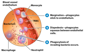
Margination: Phagocytes stick to blood vessel endothelium in response to cytokines.
Diapedesis: Phagocytes squeeze between endothelial cells to reach the site of infection.
Tissue Repair
Stroma: Supporting connective tissue that is repaired.
Parenchyma: Functioning part of the tissue that is repaired.
Fever
Fever is an abnormally high body temperature. Cytokines cause the hypothalamus to release prostaglandins, resetting the hypothalamus to a higher temperature. The body constricts blood vessels and shivers to raise temperature; vasodilation and sweating occur as temperature falls.
Antimicrobial Substances
The Complement System
The complement system consists of serum proteins produced by the liver that assist in destroying microbes. They act in a cascade, activated by pathogens or antibody-antigen reactions. Outcomes include cytolysis, opsonization, and inflammation.
Classical pathway: Antibodies bind to antigens, activating C1, which splits and activates C2 and C4, leading to C3 activation.
Alternative pathway: C3 combines with factors B, D, and P on microbe surfaces, leading to C3 activation.
Cytolysis: Membrane attack complex (MAC) forms, causing microbe lysis.
Opsonization: C3b promotes phagocyte attachment to microbes.
Inflammation: C3a binds to mast cells, releasing histamine.
Regulation: Regulatory proteins break down complement proteins to minimize host cell destruction. Lack of complement proteins increases susceptibility to infections. Capsules can prevent complement activation.
Interferons (IFNs)
IFN-α and IFN-β: Produced in response to viral infections; induce neighboring cells to produce antiviral proteins (AVPs) that inhibit viral replication.
IFN-γ: Activates neutrophils and macrophages to kill bacteria.
Iron-Binding Proteins
Transferrin: Found in blood and tissue fluids.
Lactoferrin: Found in milk, saliva, and mucus.
Ferritin: Found in liver, spleen, and red bone marrow.
Hemoglobin: Located in red blood cells.
Bacteria produce siderophores to compete with iron-binding proteins.
Antimicrobial Peptides
Short peptides that inhibit microbial growth by disrupting membranes, inhibiting cell wall synthesis, or interfering with metabolism.
