 Back
BackInnate Immunity: Nonspecific Lines of Defense in Microbiology
Study Guide - Smart Notes
 Tailored notes based on your materials, expanded with key definitions, examples, and context.
Chapter 15: Innate Immunity – Nonspecific Lines of Defense
Overview of Defense Mechanisms
The human body employs multiple layers of defense to protect against pathogenic microorganisms. These defenses are categorized as innate (nonspecific) and adaptive (specific) immunity. Innate defenses are present at birth and provide immediate, broad protection, while adaptive defenses are specific and develop after exposure to pathogens.
Innate defenses: Natural resistance to most pathogens, present before exposure.
Nonspecific defenses: First and second lines of defense, acting against a wide range of pathogens.
Specific defenses: Third line, involving lymphocytes and antibodies, targeting specific pathogens (covered in Ch. 16).
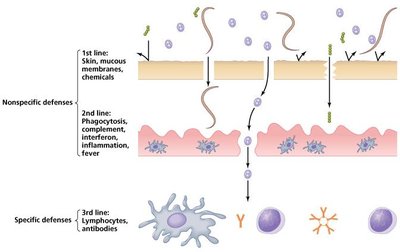
First Line of Defense: Physical and Chemical Barriers
Physical Barriers
The first line of defense consists of anatomical structures and secretions that prevent pathogen entry into the body. These barriers are nonspecific and act against a broad range of invaders.
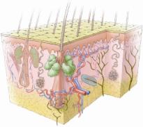
Skin: The epidermis is composed of tightly packed cells that are continually shed, removing attached microbes. Dendritic (Langerhans) cells in the epidermis perform phagocytosis.
Dermis: Contains collagen fibers for strength and sweat glands that secrete antimicrobial substances.
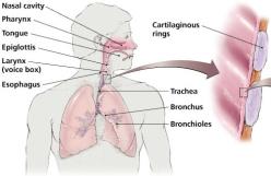
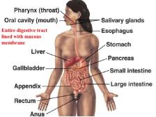
Mucous membranes: Line all body cavities open to the environment (respiratory, digestive, urinary, reproductive systems). Consist of a tightly packed epithelial layer and a supportive connective tissue layer.
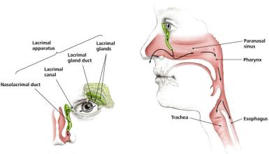
Tear ducts (lacrimal apparatus): Wash away foreign material from the eyes using fluid containing lysozyme.
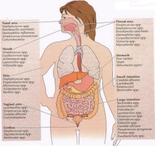
Normal flora (microbial antagonism): Compete with pathogens for nutrients and space, secrete antimicrobial substances, and alter local pH.
Mucous Membranes
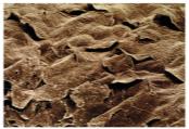
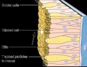
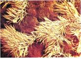
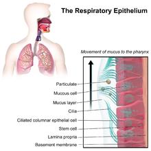
Mucous membranes are specialized to trap and remove pathogens. Goblet cells produce mucus, ciliated cells propel mucus, and dendritic cells perform phagocytosis. Lysozyme in mucus degrades bacterial cell walls.
Tear Ducts (Lacrimal Apparatus)
The lacrimal apparatus produces tears that mechanically flush the eye and contain lysozyme, which has antimicrobial properties.
Microbial Antagonism (Normal Flora)
Normal flora protect the body by outcompeting pathogens, secreting antimicrobial substances, consuming nutrients, altering pH, and stimulating the immune system. They also contribute to host health by producing vitamins (e.g., B5 in the intestine).
Chemical Barriers
Body organs secrete chemicals and enzymes that inhibit microbial growth:
Perspiration (sweat glands): Contains salt (promotes desiccation of microbes) and lysozyme (destroys bacterial cell walls).
Sebum (sebaceous glands): Keeps skin pliable and lowers pH, inhibiting many bacteria.
Other secretions: Urination, defecation, vomiting, coughing, sneezing, and crying help expel pathogens.
Antimicrobial peptides (defensins): Create holes in microbial membranes and disrupt bacterial signaling (quorum sensing).
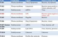
Toll-Like Receptors (TLRs): Recognize common microbial molecules and trigger immune responses.
Second Line of Defense: Internal Nonspecific Responses
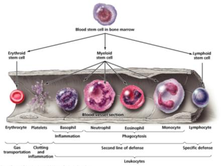
Blood and Immune Cells
When pathogens breach the first line of defense, the second line is activated. This includes cellular and chemical components found in blood, but not physical barriers.
Plasma: Contains water, electrolytes, nutrients, proteins (including complement proteins and antibodies).
Serum: Plasma without clotting factors.
Formed elements: Erythrocytes (gas transport), platelets (clotting), leukocytes (defense).
Leukocytes: Granulocytes (basophils, eosinophils, neutrophils) and agranulocytes (lymphocytes, monocytes).
Phagocytosis
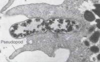
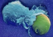
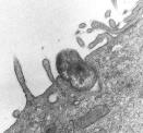
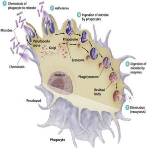
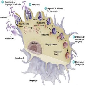
Phagocytes (certain leukocytes and derivatives) ingest and destroy pathogens in a tightly regulated process. The five stages are:
Chemotaxis: Movement toward chemical signals (cytokines).
Adherence: Attachment to microbes via receptors (opsonization enhances this step).
Ingestion: Formation of a phagosome (vacuole).
Digestion: Fusion of phagosome with lysosome; enzymes degrade the microbe.
Elimination: Expulsion of digested material.
Phagocytosis – Blocking Mechanisms
Some pathogens evade phagocytosis by various mechanisms, such as preventing chemotaxis, adherence, ingestion, or digestion.
Protection of Self
Body cells are marked with autoantigens (MHC) to prevent phagocytosis. Phagocytes have receptors for microbial components not found on host cells, and signaling molecules (complement, antibodies, interferons, defensins) provide targeted instructions.
Inflammation
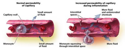
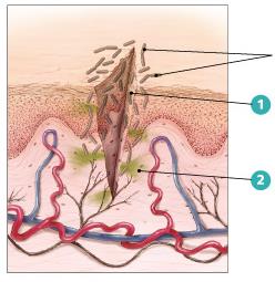
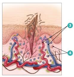
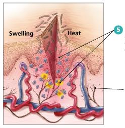
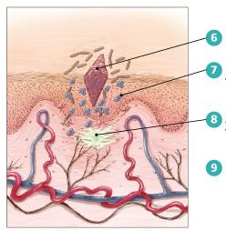
Inflammation is a nonspecific response to tissue damage, characterized by redness, heat, swelling, and pain. It can be acute (short-lived, beneficial) or chronic (long-lasting, potentially damaging).
Acute inflammation: Involves vasodilation, increased permeability, migration of phagocytes, and tissue repair.
Chronic inflammation: Prolonged, can damage tissues.
Fever
Fever is an elevated body temperature triggered by pyrogens (e.g., bacterial toxins, interleukin-1). It enhances phagocyte activity, tissue repair, and interferon effects, while inhibiting some pathogens.
Nonspecific Chemical Defenses
Chemical responses protect against broad classes of invaders and promote phagocytosis, inflammation, and fever. Major systems include:
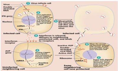
Interferon system: Proteins released by virus-infected cells signal neighboring cells to produce antiviral proteins (AVPs), which block viral replication.
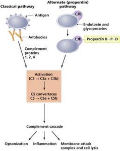
Complement system: Serum proteins activated by infection that assist in phagocytosis (opsonization), promote inflammation, and form membrane attack complexes (MAC) to lyse foreign cells.
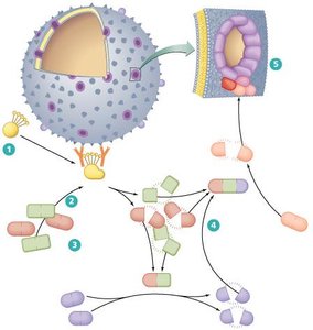
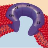
Summary Table: Main Functions of the Complement System
Function | Description |
|---|---|
Opsonization | Coating of microbes to enhance phagocytosis |
Promotion of Inflammation | Recruitment and activation of immune cells |
Membrane Attack Complex (MAC) | Formation of pores in pathogen membranes, leading to lysis |
Key Terms and Concepts
Innate immunity: Nonspecific, immediate defense present at birth.
Phagocytosis: Cellular process of engulfing and destroying pathogens.
Inflammation: Localized response to tissue injury or infection.
Fever: Systemic increase in body temperature as a defense mechanism.
Interferons: Proteins that inhibit viral replication.
Complement system: Group of proteins that enhance immune responses.
Normal flora: Beneficial microorganisms that inhabit the body and prevent pathogen colonization.
Equations and Formulas
Fever temperature conversion:
Additional info: This guide covers the main concepts of innate immunity, focusing on the first and second lines of defense as outlined in Chapter 15 of a typical microbiology curriculum. For adaptive (specific) immunity, see Chapter 16.
