Back
BackInnate Immunity: Principles and Clinical Applications
Study Guide - Smart Notes
 Tailored notes based on your materials, expanded with key definitions, examples, and context.
Innate Immunity: Principles and Clinical Applications
Overview of the Immune System
The immune system is a complex network that protects the body from pathogens. It is divided into two main branches: innate immunity and adaptive immunity. Both branches share the ability to recognize and eliminate diverse pathogens while distinguishing self from non-self antigens.
Innate Immunity: Inborn, non-specific, and present in all eukaryotes. Provides immediate defense against infection.
Adaptive Immunity: Found only in vertebrates, matures over time, and tailors responses to specific pathogens. Exhibits immunological memory.
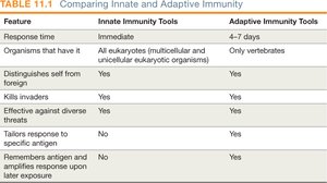
Table Purpose: Comparison of innate and adaptive immunity features, including response time, specificity, and memory.
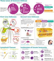
Three Lines of Immune Defense
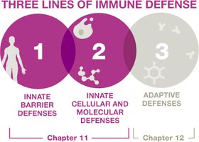
Immunity is organized into three lines of defense:
First Line: Innate barrier defenses (physical, mechanical, chemical barriers)
Second Line: Innate cellular and molecular defenses (leukocytes, inflammation, fever, complement system)
Third Line: Adaptive defenses (lymphocytes, antibodies)
Normal Microbiota and Immune Responses
Role of Normal Microbiota
Normal microbiota are the diverse microbes that colonize the human body. They play a crucial role in shaping, training, and calibrating immune responses. Disruptions in microbiota composition can lead to immune confusion, potentially contributing to allergies and autoimmune diseases (hygiene hypothesis).
Germ-free animal studies show underdeveloped immune systems, highlighting the importance of microbiota in immune development.
First-Line Defenses
Barriers to Pathogen Entry
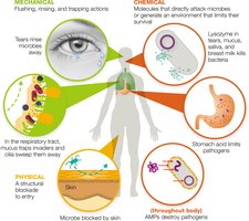
First-line defenses aim to prevent pathogen entry and are categorized as mechanical, chemical, and physical barriers.
Mechanical Barriers: Actions that rinse, flush, or trap pathogens (e.g., tears, urine, saliva, mucus, mucociliary escalator).
Chemical Barriers: Molecules that attack microbes or create hostile environments (e.g., lysozyme in tears and saliva, stomach acid, fatty acids in sweat and earwax).
Physical Barriers: Structures that physically block entry (e.g., skin, tightly packed epithelial cells).
Antimicrobial Peptides (AMPs)
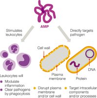
AMPs are proteins that destroy a wide range of pathogens. Defensins are a key class of AMPs that disrupt microbial membranes.
Second-Line Defenses and the Lymphatic System
Lymphatic System Function
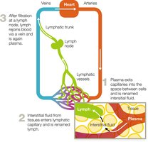
The lymphatic system collects, circulates, and filters body fluids, playing a central role in immune surveillance and response. Lymph is interstitial fluid collected by lymphatic capillaries, filtered through lymph nodes, and returned to the bloodstream.
Primary and Secondary Lymphoid Tissues
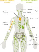
Primary Lymphoid Tissues: Sites of leukocyte production and maturation (bone marrow, thymus).
Secondary Lymphoid Tissues: Sites where immune responses are initiated (lymph nodes, spleen, MALT).
Leukocytes and Immune Responses
Types of Leukocytes
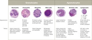
Leukocytes (white blood cells) are essential for immune responses and are classified as granulocytes or agranulocytes.
Granulocytes: Neutrophils, eosinophils, basophils, mast cells
Agranulocytes: Monocytes (macrophages), dendritic cells, lymphocytes (NK cells, B cells, T cells)
Clinical Relevance: Leukocytosis
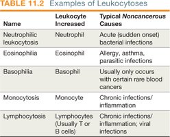
White blood cell counts are used clinically to diagnose infections and immune disorders. Increases in specific leukocyte types can indicate particular conditions.
Name | Leukocyte Increased | Typical Noncancerous Causes |
|---|---|---|
Neutrophilic leukocytosis | Neutrophil | Acute bacterial infections |
Eosinophilia | Eosinophil | Allergy, asthma, parasitic infections |
Basophilia | Basophil | Rare blood cancers |
Monocytosis | Monocyte | Chronic infections/inflammation |
Lymphocytosis | Lymphocytes | Chronic infections/inflammation; viral infections |
Cellular Second-Line Defenses
Phagocytes and Their Functions
Phagocytes (macrophages, dendritic cells, neutrophils) engulf and destroy pathogens. Some microbes possess virulence factors to evade phagocytosis.
Neutrophils: Most abundant, first responders, release AMPs, phagocytize pathogens.
Eosinophils: Combat parasites, moderate phagocytic activity, involved in allergies.
Basophils and Mast Cells: Release histamine, involved in allergic responses and fighting parasites.
Monocytes/Macrophages: Largest WBCs, mature into macrophages in tissues, highly phagocytic.
Dendritic Cells: Patrol tissues, present antigens to adaptive immune cells.
Lymphocytes: NK cells (innate), B and T cells (adaptive).
Molecular Second-Line Defenses
Key Molecular Defenses
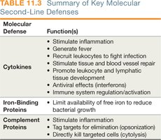
Second-line molecular defenses include cytokines, iron-binding proteins, and complement proteins.
Molecular Defense | Function(s) |
|---|---|
Cytokines | Stimulate inflammation, fever, recruit leukocytes, tissue repair, antiviral effects, immune regulation |
Iron-Binding Proteins | Limit free iron to reduce bacterial growth |
Complement Proteins | Stimulate inflammation, opsonization, cytolysis |
Cytokines
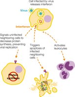
Cytokines are signaling proteins that coordinate immune responses. Major classes include:
Chemokines: Attract leukocytes to infection sites (chemotaxis).
Interleukins (ILs): Regulate inflammation, hematopoiesis, and immune responses.
Interferons (IFNs): Antiviral signaling, activate immune cells.
Tumor Necrosis Factors (TNFs): Stimulate inflammation, fever, and kill tumor cells.
Iron-Binding Proteins
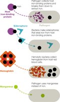
Iron is essential for microbial growth. Host proteins (hemoglobin, ferritin, lactoferrin, transferrin) sequester iron to limit pathogen access. Some pathogens produce siderophores to steal iron or lyse red blood cells to access hemoglobin.
Complement System
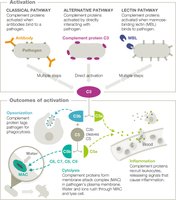
The complement system is a cascade of >30 proteins that enhance immune responses. Activation occurs via three pathways:
Classical Pathway: Triggered by antibodies bound to pathogens.
Alternative Pathway: Direct interaction with pathogen surfaces.
Lectin Pathway: Initiated by mannose-binding lectin binding to microbial sugars.
All pathways result in:
Opsonization: Tagging pathogens for phagocytosis
Membrane Attack Complex (MAC): Cytolysis of target cells
Inflammation: Recruitment of immune cells
Inflammation and Fever
Inflammation
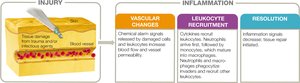
Inflammation is a protective response to tissue injury or infection, essential for healing but potentially damaging if unregulated. It occurs in three phases:
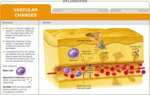
Vascular Changes: Vasodilation and increased permeability allow immune cells and proteins to access tissues.
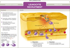
Leukocyte Recruitment: Chemotaxis brings leukocytes to the site; neutrophils arrive first, followed by monocytes.
Resolution: Inflammation subsides, tissue repair begins, and pus may form.
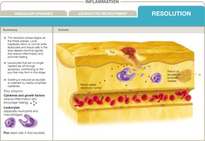
Cardinal Signs of Inflammation: Redness, pain, localized heat, swelling, loss of function.
Key Chemical Mediators: Histamine, kinins, eicosanoids (prostaglandins, leukotrienes, thromboxanes).
Fever
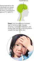
Fever (pyrexia) is a systemic response to infection, mediated by pyrogens (e.g., bacterial toxins, cytokines) that signal the hypothalamus to raise body temperature. Fever enhances immune efficiency but can be dangerous if too high.
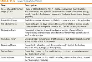
Term | Description |
|---|---|
Fever of undetermined origin (FUO) | Fever >38.3°C (101°F) for >3 weeks without a clear cause |
Intermittent fever | Body temperature elevates, then falls to normal |
Remittent fever | Elevated temperature fluctuates, but doesn't reach normal |
Sustained fever | Constantly elevated temperature with minimal fluctuation |
Tertian/Quartian fever | Fever patterns seen in malaria |
Clinical Application: Case Study
Case Summary
A 5-year-old boy with recurrent infections was diagnosed with bacterial meningitis caused by Streptococcus pneumoniae. Despite vaccination, he developed infection due to a strain not covered by the vaccine. Further investigation revealed a deficiency in mannose-binding lectin (MBL), affecting his lectin pathway of complement activation.
Key Points: MBL deficiency increases susceptibility to infections; adaptive immunity (e.g., vaccine response) remains functional.
Clinical Measures: Differential WBC count, assessment for immune deficiencies, and iron metabolism disorders (e.g., hemochromatosis).
Visual Summary