 Back
BackInnate Immunity: Principles and Mechanisms
Study Guide - Smart Notes
 Tailored notes based on your materials, expanded with key definitions, examples, and context.
Innate Immunity
Overview of Immune Responses
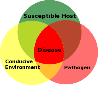
The immune system coordinates physiological processes to eliminate antigens and protect the host from infection. Immunity can be classified as innate (inborn, nonspecific) or adaptive (acquired, specific). Both systems recognize diverse pathogens, eliminate invaders, and distinguish self from non-self antigens.
Immune: Specific protection conferred by immune responses.
Susceptible: Not immune to a given pathogen and may develop infection.
Normal microbiota play a central role in training and calibrating immune responses. The good hygiene hypothesis suggests that reduced microbial exposure may impair immune system development.
Lines of Immune Defense
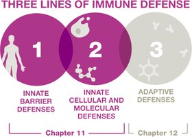
The immune system is organized into three lines of defense:
First-line defenses: Innate barriers that prevent pathogen entry (mechanical, chemical, and physical barriers).
Second-line defenses: Innate cellular and molecular defenses (e.g., leukocytes, antimicrobial proteins).
Third-line defenses: Adaptive immune responses (T and B lymphocytes).
First-line Defenses
Mechanical Barriers
Mechanical barriers physically remove pathogens from body surfaces through rinsing, flushing, or trapping actions.
Tears wash pathogens from eyes.
Urine flushes microbes from the urinary tract.
Saliva limits microbial adherence in the mouth.
Mucus membranes trap microbes; the mucociliary escalator sweeps them out of the respiratory tract.

Chemical Barriers
Chemical barriers directly attack invaders or create hostile environments for pathogens.
Lysozyme: Enzyme in tears, saliva, and breast milk that breaks down bacterial cell walls.
Hydrochloric acid: Stomach acid that destroys ingested microbes.
Skin: Dry, salty, and slightly acidic, inhibiting microbial growth.
Fatty acids: Present in sweat and earwax, providing antimicrobial activity.
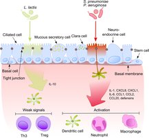
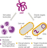
Antimicrobial peptides (AMPs): Proteins such as defensins that destroy a wide range of pathogens.
Physical Barriers
Physical barriers are anatomical structures that block pathogen entry.
Skin: The epidermis consists of tightly packed dead epithelial cells, forming a robust barrier.
Second-line and Third-line Defenses
When pathogens bypass first-line defenses, they encounter second-line defenses (molecular factors and leukocytes) and third-line defenses (adaptive immunity mediated by T and B lymphocytes).
Organs, Tissues, Cells, and Molecules of the Immune System
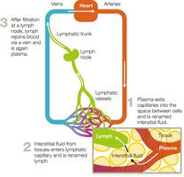
Lymphatic System
The lymphatic system is essential for immune function, collecting, circulating, and filtering tissue fluid before returning it to the bloodstream.
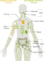
Primary and Secondary Lymphoid Tissues
Primary lymphoid tissues: Sites of leukocyte production and maturation (thymus and bone marrow).
Secondary lymphoid tissues: Sites where immune responses are initiated (lymph nodes, spleen, MALT).
Blood and Leukocytes
Blood contains erythrocytes (oxygen transport), platelets (clotting), and leukocytes (immune defense). Leukocytes are classified as granulocytes or agranulocytes based on cytoplasmic granules.
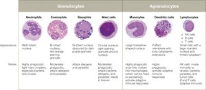
Granulocytes
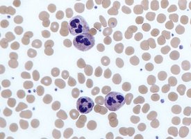
Neutrophils: Most abundant WBCs, first responders to infection, phagocytic, release antimicrobial peptides.
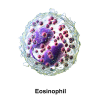
Eosinophils: Combat parasites, moderate phagocytic activity, granules contain enzymes and toxins.
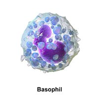
Basophils: Involved in allergic responses and parasitic infections, granules contain histamine.
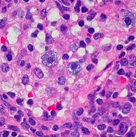
Mast cells: Tissue-resident, release histamine, involved in allergies and parasite defense, capable of phagocytosis.
Agranulocytes
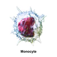
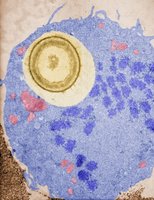
Monocytes: Largest WBCs, migrate into tissues to become macrophages, elevated in chronic infections.
Macrophages: Highly phagocytic, can be fixed (tissue-resident) or wandering.
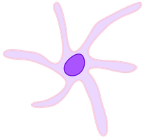
Dendritic cells: Highly phagocytic, patrol tissues, present antigens to adaptive immune cells.
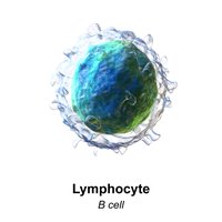
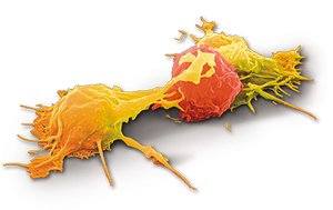
Lymphocytes: Include natural killer (NK) cells (innate immunity), B cells, and T cells (adaptive immunity).
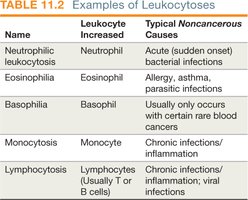
White Blood Cell Counts in Clinical Diagnosis
Differential WBC counts are used to diagnose infections and immune disorders. Leukocytosis is an increase, and leukopenia is a decrease in WBCs.
Immune Molecules
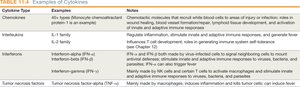
Cytokines: Signaling proteins for cell communication and immune coordination.
Chemokines: Induce chemotaxis, recruit cells to infection sites, aid in tissue repair.
Interleukins (ILs): Activate immune responses, regulate inflammation, stimulate hematopoiesis.
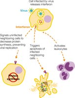
Interferons (IFNs): Signal viral infection, induce antiviral defenses, activate leukocytes.
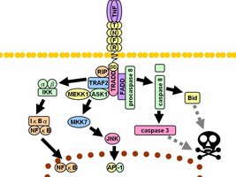
Tumor necrosis factors (TNFs): Stimulate inflammation, fever, and can kill tumor cells.
Iron-Binding Proteins and Pathogen Strategies
Iron is sequestered by proteins such as hemoglobin, ferritin, lactoferrin, and transferrin to limit microbial growth. Some pathogens produce siderophores to steal iron or lyse red blood cells to access hemoglobin.
Complement System
The complement system consists of over 30 proteins that, when activated, enhance immune defenses through a cascade mechanism. The three main outcomes are:
Opsonization: Tagging invaders for phagocytosis.
Membrane Attack Complex (MAC) formation: Causing cytolysis of pathogens.
Inflammation: Promoting recruitment of immune cells and enhancing responses.
Inflammation
Phases and Goals of Inflammation
Inflammation is a key innate defense mechanism triggered by tissue damage or infection. Its goals are to recruit immune defenses, limit pathogen spread, and promote tissue repair.
Cardinal signs: Redness, pain, localized heat, swelling, loss of function.
Phases of Inflammation
Vascular changes: Vasodilation and increased vessel permeability allow immune cells and proteins to access tissues.
Leukocyte recruitment: Chemoattractants guide leukocytes to the site; margination and diapedesis facilitate their exit from blood vessels.
Resolution: Signals tone down inflammation, clear exudate, and initiate tissue repair.
Inflammatory Mediators
Histamine: Released by mast cells, basophils, eosinophils, and platelets; increases vessel permeability.
Kinins: Induce vascular changes and pain, assist in clotting.
Eicosanoids: Prostaglandins, leukotrienes, and thromboxanes; mediate inflammation and pain.
Chronic Inflammation
Chronic inflammation results from prolonged immune activation, causing tissue injury and contributing to diseases such as atherosclerosis, cancer, and neurodegeneration.
Fever (Pyrexia)
Mechanisms and Effects
Fever is an abnormally high systemic body temperature, induced by pyrogens (e.g., bacterial toxins, cytokines) that signal the hypothalamus to raise the body's set point.
Low-grade fever (37.5°C to 38.3°C): Generally protective, enhances interferon activity, phagocytosis, leukocyte production, and tissue repair.
High fever (≥40.5°C): Life-threatening, can denature proteins and enzymes, and is a medical emergency.
Antipyretics (e.g., aspirin, ibuprofen, acetaminophen) are used to lower fever by inhibiting prostaglandin production in the hypothalamus.
Summary Table: Leukocyte Types and Functions
Leukocyte | Granules | Function |
|---|---|---|
Neutrophil | Yes | Phagocytosis, first responder to infection |
Eosinophil | Yes | Combat parasites, moderate phagocytosis |
Basophil | Yes | Release histamine, allergic responses |
Mast cell | Yes | Release histamine, tissue resident |
Monocyte | No | Differentiate into macrophages, phagocytosis |
Macrophage | No | Phagocytosis, antigen presentation |
Dendritic cell | No | Phagocytosis, antigen presentation |
Lymphocyte (NK, B, T) | No | NK: innate cytotoxicity; B/T: adaptive immunity |
Summary Table: Examples of Cytokines
Cytokine Type | Examples | Notes |
|---|---|---|
Chemokines | Monocyte chemoattractant protein-1 | Induce chemotaxis, wound healing, lymphoid tissue development |
Interleukins | IL-1, IL-2 | Activate immune responses, regulate inflammation, fever, apoptosis |
Interferons | IFN-α, IFN-β, IFN-γ | Antiviral responses, activate leukocytes |
Tumor necrosis factor | TNF-α | Stimulates inflammation, fever, kills tumor cells |
