 Back
BackInnate Immunity: The Body’s First and Second Lines of Defense
Study Guide - Smart Notes
 Tailored notes based on your materials, expanded with key definitions, examples, and context.
Innate Immunity: The Body’s First and Second Lines of Defense
Overview of Host Defenses
Innate immunity provides the body’s immediate, nonspecific defense against pathogens. It consists of physical barriers, cellular responses, and chemical mediators that act rapidly to prevent and control infections.
The Body’s First Line of Defense
Physical and Chemical Barriers
Skin and mucous membranes are the primary barriers preventing pathogen entry.
These barriers are supported by antimicrobial chemicals and physiological processes.
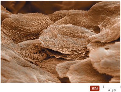
The Role of Skin in Innate Immunity
The epidermis consists of multiple layers of tightly packed cells, making it difficult for pathogens to penetrate. Shedding of dead skin cells removes attached microorganisms.
Epidermal dendritic cells phagocytize pathogens.
The dermis contains collagen fibers that provide resistance to abrasions.
Skin secretions include:
Perspiration: Contains salt (inhibits microbial growth), antimicrobial peptides, and lysozyme (destroys bacterial cell walls).
Sebum: Maintains pliability and lowers skin pH, inhibiting many bacteria.
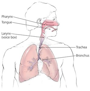
The Role of Mucous Membranes in Innate Immunity
Line all body cavities open to the environment (respiratory, digestive, urinary, reproductive tracts).
Composed of:
Epithelium: Thin, living, tightly packed cells that are continually shed, removing microbes. Goblet and ciliated cells help trap and remove invaders.
Deeper connective tissue: Supports the epithelium and produces antimicrobial chemicals.
Comparison of Skin and Mucous Membranes
Feature | Skin | Mucous Membrane |
|---|---|---|
Number of Cell Layers | Many | One to a few |
Cells Tightly Packed? | Yes | Yes |
Cells Dead or Alive? | Outer: dead; Inner: alive | Alive |
Mucus Present? | No | Yes |
Lysozyme Present? | Yes | With some |
Sebum Present? | Yes | No |
Cilia Present? | No | Trachea, uterine tubes |
Constant Shedding? | Yes | Yes |
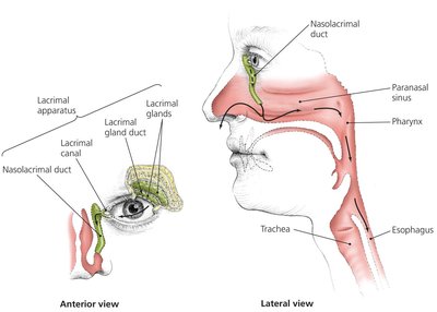
The Role of the Lacrimal Apparatus
Produces and drains tears, washing the surface of the eye.
Tears contain lysozyme, which destroys bacteria.
The Role of the Microbiome
Microbial antagonism: Normal microbiota compete with pathogens for nutrients and attachment sites, create unfavorable environments, and stimulate the immune system.
Some microbiota produce antimicrobial compounds and vitamins, and modulate immunity.
Other First-Line Defenses
Antimicrobial peptides: Present in skin, mucous membranes, and neutrophils; act against a variety of microbes.
Other organs secrete chemicals with antimicrobial properties (e.g., saliva, stomach acid, bile, urine, vaginal secretions).
Secretions and Activities Contributing to the First Line of Defense
System | Secretion/Activity | Function |
|---|---|---|
Digestive | Saliva | Washes microbes, contains lysozyme |
Digestive | Stomach acid | Digests/inhibits microorganisms |
Digestive | Bile | Inhibitory to most microorganisms |
Urinary | Urine | Acidity inhibits microbes, washes ureters/urethra |
Reproductive | Vaginal secretions | Acidity inhibits microbes, sequesters iron |
Cardiovascular | Blood flow | Removes microbes from wounds |
The Body’s Second Line of Defense
Defense Components of Blood
Plasma: Contains water, electrolytes, proteins (including complement and antibodies), and iron-binding compounds.
Formed elements:
Erythrocytes: Transport oxygen and carbon dioxide.
Platelets: Involved in clotting and inflammation.
Leukocytes: Defend against invaders; divided into granulocytes and agranulocytes.
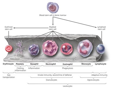
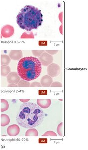
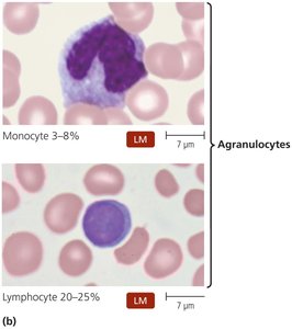
Leukocytes: Granulocytes and Agranulocytes
Granulocytes:
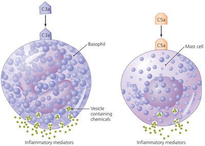
Basophils: Release inflammatory chemicals.
Eosinophils: Phagocytize pathogens, attack helminths, involved in allergies.
Neutrophils: Phagocytize pathogens, can kill without phagocytosis.
Agranulocytes:
Lymphocytes: Most involved in adaptive immunity; natural killer (NK) cells kill infected/tumor cells.
Monocytes: Mature into macrophages, which are phagocytic.
Lab Analysis of Leukocytes
Differential white blood cell counts can indicate disease:
Increased eosinophils: Allergies or parasitic infection
Increased neutrophils: Bacterial infection
Increased lymphocytes: Viral infection
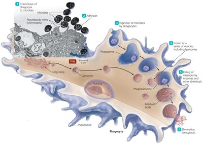
Phagocytosis
Phagocytosis is the process by which certain cells (phagocytes) ingest and destroy pathogens. It occurs in six stages:
Chemotaxis
Adhesion
Ingestion
Maturation
Killing
Elimination
Nonphagocytic Killing
Eosinophils: Secrete toxins to kill helminths; release mitochondrial DNA/proteins to kill bacteria.
Natural Killer (NK) cells: Secrete toxins onto virally infected or tumor cells.
Neutrophils: Release chemicals and form neutrophil extracellular traps (NETs) to kill microbes.
Nonspecific Chemical Defenses
Toll-like receptors (TLRs): Recognize pathogen-associated molecular patterns (PAMPs) and trigger immune responses (e.g., inflammation, apoptosis).
NOD proteins: Cytosolic proteins that bind PAMPs and trigger innate responses.
Interferons: Proteins released by host cells to inhibit viral spread; include Type I (alpha, beta) and Type II (gamma) interferons.
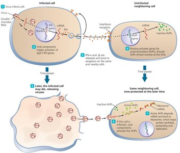
Table: Characteristics of Human Interferons
Type | Principal Source | Inducing Agent | Action |
|---|---|---|---|
Alpha (IFN-α) | Epithelium, leukocytes | Viruses | Stimulates production of antiviral proteins |
Beta (IFN-β) | Fibroblasts | Viruses | Stimulates production of antiviral proteins |
Gamma (IFN-γ) | Activated T and NK lymphocytes | Adaptive immune responses | Stimulates phagocytic activity |
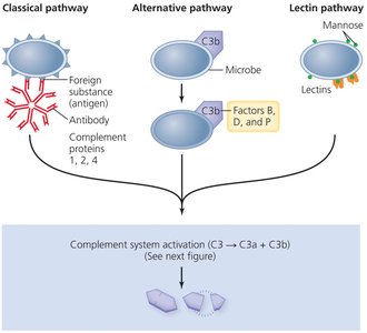
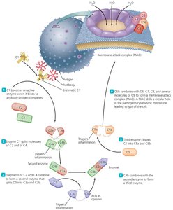
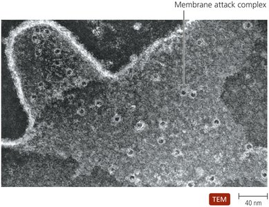
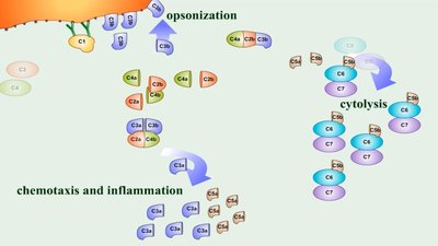
Complement System
A set of serum proteins that, when activated, result in lysis of foreign cells, inflammation, and opsonization.
Activation pathways: Classical, alternative, and lectin.
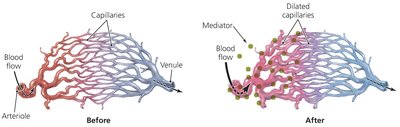
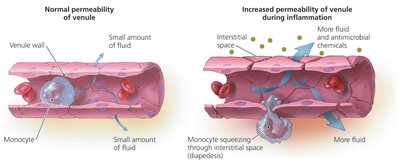
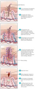
Inflammation
A nonspecific response to tissue damage, characterized by redness, heat, swelling, and pain.
Types:
Acute: Rapid, short-lived, beneficial for defense and repair.
Chronic: Long-lasting, can cause tissue damage.
Key events:
Vasodilation and increased vascular permeability (mediated by histamine, prostaglandins, etc.)
Migration of phagocytes to the site of infection
Tissue repair
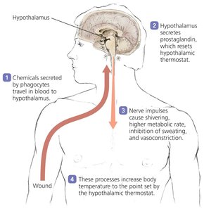
Fever
Defined as a body temperature above 37°C, triggered by pyrogens acting on the hypothalamus.
Pyrogens include bacterial toxins, cytoplasmic contents of bacteria, antibody-antigen complexes, and pyrogens released by phagocytes.
Fever enhances interferon effects, inhibits some microbes, and may enhance phagocyte and tissue repair activities.
Summary Table: Nonspecific Components of Innate Immunity
First Line | Second Line |
|---|---|
Barriers (skin, mucous membranes, chemicals) | Phagocytes (macrophages, neutrophils, eosinophils) |
Antimicrobial peptides | Extracellular killing (eosinophils, NK cells, neutrophils) |
Secretions (sweat, acid, lysozyme, mucus) | Complement, interferons, inflammation, fever |
Key Terms
Innate immunity: Nonspecific defense mechanisms present from birth.
Phagocytosis: Cellular process of engulfing and destroying pathogens.
Complement system: Group of proteins that enhance immune responses.
Inflammation: Localized response to injury or infection.
Fever: Systemic increase in body temperature as a defense mechanism.
