 Back
BackInnate Immunity: The Body’s First and Second Lines of Defense
Study Guide - Smart Notes
 Tailored notes based on your materials, expanded with key definitions, examples, and context.
An Overview of the Body’s Defenses
Introduction to Host Defenses
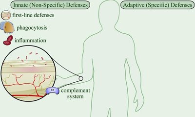
The human body is equipped with multiple defense mechanisms to resist infection by pathogens. These defenses are categorized into innate (non-specific) and adaptive (specific) immunity. Innate immunity provides immediate, general protection, while adaptive immunity targets specific pathogens after exposure.
Species resistance: Humans are naturally resistant to many pathogens due to physiological differences, such as the absence of specific receptors or incompatible environmental conditions for pathogen survival.
First-line defenses: Physical and chemical barriers that prevent pathogen entry.
Second-line defenses: Cellular and chemical responses activated if pathogens breach the first line.
The Body’s First Line of Defense
Physical and Chemical Barriers
The first line of defense consists of structures, chemicals, and processes that prevent pathogens from entering the body. The primary components are the skin and mucous membranes, which cover all body surfaces exposed to the environment.
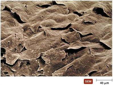
Skin: Composed of the epidermis (multiple layers of tightly packed cells) and dermis (collagen fibers for strength). The shedding of dead skin cells removes attached microorganisms, and dendritic cells in the epidermis phagocytize invaders.
Chemical defenses of skin:
Perspiration (sweat) contains salt (inhibits microbial growth), antimicrobial peptides, and lysozyme (destroys bacterial cell walls).
Sebum (oil) keeps skin pliable and lowers pH, inhibiting many bacteria.
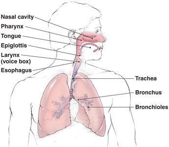
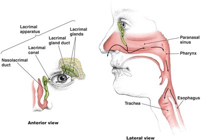
Mucous Membranes and the Lacrimal Apparatus
Mucous membranes line all body cavities open to the environment, such as the respiratory, digestive, urinary, and reproductive tracts. They consist of a thin epithelium and a deeper connective tissue layer.
Epithelium: Tightly packed, living cells that are continually shed, removing microorganisms.
Lacrimal apparatus: Produces and drains tears, which contain lysozyme to destroy bacteria and physically wash the eye surface.
The Role of Normal Microbiota
Normal microbiota (resident microbes) protect the host through microbial antagonism, competing with potential pathogens for nutrients and space, and producing substances that inhibit pathogen growth. They also stimulate the body’s second line of defense and contribute to host health by synthesizing vitamins.
Other First-Line Defenses
Antimicrobial peptides: Present in skin, mucous membranes, and neutrophils; act against a variety of microbes by disrupting membranes or interfering with metabolism.
Other chemicals: Many organs secrete antimicrobial substances as part of their protective function.
The Body’s Second Line of Defense
Defense Components of Blood
When pathogens penetrate the first line of defense, the second line is activated. This includes cellular and chemical components, many of which are found in the blood.
Plasma: The liquid portion of blood, containing water, electrolytes, nutrients, proteins (including complement proteins and antibodies), and iron-binding compounds.
Formed elements:
Erythrocytes: Transport oxygen and carbon dioxide.
Platelets: Involved in blood clotting and inflammation.
Leukocytes (white blood cells): Defend against invaders and are divided into granulocytes and agranulocytes.
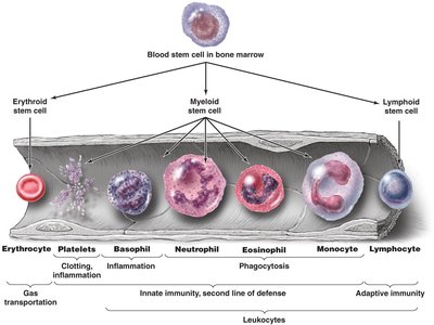
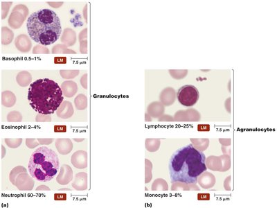
Granulocytes and Agranulocytes
Granulocytes: Contain visible granules; include basophils (inflammation), eosinophils (phagocytosis, defense against parasites), and neutrophils (phagocytosis, most abundant).
Agranulocytes: Lack visible granules; include lymphocytes (adaptive immunity) and monocytes (mature into macrophages).
Lab analysis of leukocyte counts can indicate infection type: increased eosinophils suggest parasitic infection, increased neutrophils suggest bacterial infection, and increased lymphocytes suggest viral infection.
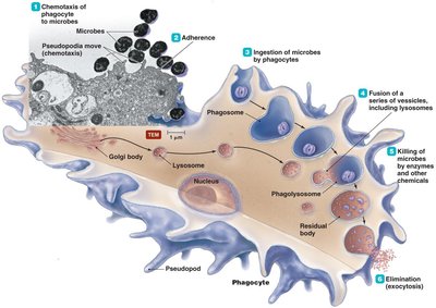
Phagocytosis
Phagocytosis is the process by which certain cells (phagocytes) ingest and destroy pathogens. It occurs in six stages:
Chemotaxis: Movement of phagocytes toward chemical signals from pathogens or damaged cells.
Adherence: Attachment of phagocyte to microbe, often enhanced by opsonins (antibody or complement proteins).
Ingestion: Engulfment of the microbe into a phagosome.
Maturation: Fusion of the phagosome with lysosomes to form a phagolysosome.
Killing: Destruction of the microbe by enzymes and toxic substances.
Elimination: Expulsion of microbial debris by exocytosis.
Nonphagocytic Killing
Eosinophils: Attack parasitic worms by secreting toxins; can also kill some bacteria with mitochondrial DNA and proteins.
Natural killer (NK) lymphocytes: Secrete toxins onto the surface of virally infected cells and tumors, sparing normal cells.
Neutrophils: Release chemicals and form neutrophil extracellular traps (NETs) to bind and kill bacteria.
Nonspecific Chemical Defenses Against Pathogens
Toll-like receptors (TLRs): Membrane proteins on phagocytes that recognize pathogen-associated molecular patterns (PAMPs) and trigger defensive responses such as apoptosis, inflammation, or stimulation of adaptive immunity.
NOD proteins: Cytosolic proteins that detect PAMPs inside the cell and initiate immune responses.
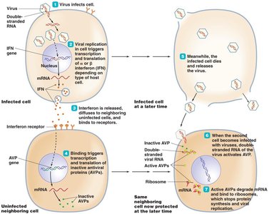
Interferons: Proteins released by host cells to inhibit viral replication.
Type I (alpha and beta): Induce antiviral proteins in neighboring cells.
Type II (gamma): Activates macrophages and enhances adaptive immunity.
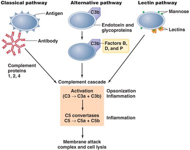
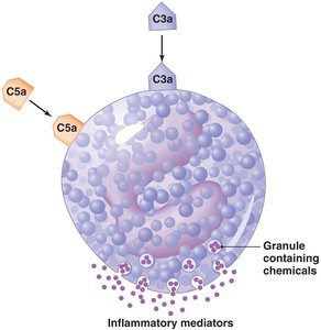
Complement System
The complement system is a group of serum proteins that, when activated, enhance the ability of antibodies and phagocytic cells to clear microbes and damaged cells. Activation leads to cell lysis, inflammation, and opsonization. There are three activation pathways:
Classical pathway: Triggered by antibodies bound to antigens.
Alternative pathway: Triggered by microbial surfaces.
Lectin pathway: Triggered by mannose-binding lectin binding to pathogen surfaces.
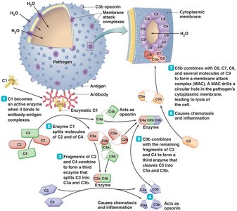
Body cells have mechanisms to inactivate complement proteins, preventing damage to host tissues.
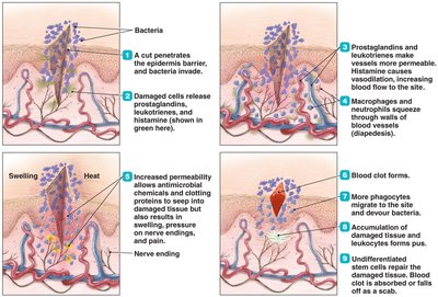
Inflammation
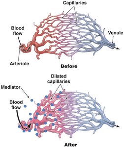
Inflammation is a nonspecific response to tissue damage, characterized by redness, heat, swelling, and pain. It can be acute (short-lived and beneficial) or chronic (long-lasting and potentially harmful).
Acute inflammation:
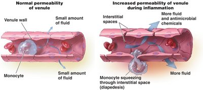
Vasodilation and increased vascular permeability allow immune cells and antimicrobial chemicals to reach the site of injury.
Phagocytes migrate to the site and remove pathogens.
Tissue repair follows pathogen elimination.
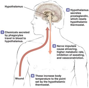
Fever
Fever is an elevated body temperature above 37°C, triggered by pyrogens (such as bacterial toxins, cytoplasmic contents of bacteria, or antibody-antigen complexes) that signal the hypothalamus to increase the body’s temperature set point. Fever enhances the effects of interferons, inhibits some microbial growth, and may enhance tissue repair.
Additional info: The content above is based on the principles of innate immunity as covered in standard microbiology textbooks, specifically focusing on the body's first and second lines of defense (Ch. 15 - Innate Immunity). Where the original material was brief, academic context and definitions were expanded for clarity and completeness.
