 Back
BackInnate Immunity: The Body’s First and Second Lines of Defense
Study Guide - Smart Notes
 Tailored notes based on your materials, expanded with key definitions, examples, and context.
Innate Immunity
Definitions and Overview
Innate immunity refers to the non-specific defense mechanisms that are present from birth and provide the first line of defense against pathogens. These responses are rapid, do not target specific pathogens, and do not generate immunological memory.
Immunity: Resistance to infection or disease.
Susceptibility: Lack of immunity, leading to vulnerability to infection.
Innate Immunity: Always present, immediate, non-specific, and without memory.
Goal: Prevent microbes from gaining access to the body.
First Line of Defense
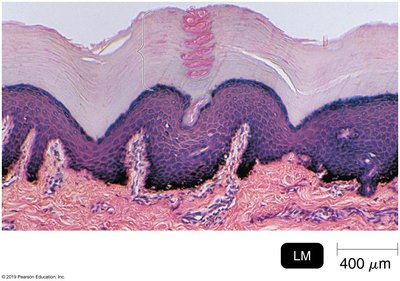
Skin
The skin acts as a large physical barrier to microbial invasion, consisting of two main layers:
Epidermis: The outer, thinner layer composed of multiple layers of epithelial cells containing keratin, which provides toughness and water-resistance.
Dermis: The inner, thicker layer made of connective tissue.
Protective Mechanisms: Shedding of epidermal cells, dryness, and secretion of oils and chemicals help prevent microbial colonization.
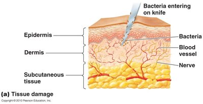
Subcutaneous Infections: Occur when the outer layer is penetrated, allowing microbes to enter underlying tissues.
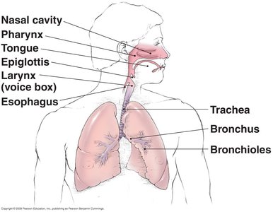
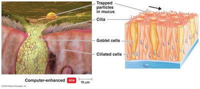
Mucous Membranes
Mucous membranes line body cavities that are open to the environment and provide both physical and chemical barriers to pathogens.
Components: Epithelial layer with goblet cells (secrete mucus) and ciliated cells (move mucus), supported by underlying connective tissue.
Locations: Gastrointestinal, genitourinary, respiratory tracts, and conjunctiva of the eyes.
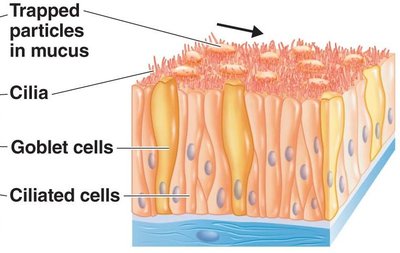
Ciliary Escalator
The ciliary escalator is a mechanism in the respiratory tract where cilia move mucus (containing trapped particles and microbes) toward the throat, where it can be swallowed and destroyed in the stomach.
Function: Traps and removes inhaled particles and pathogens.
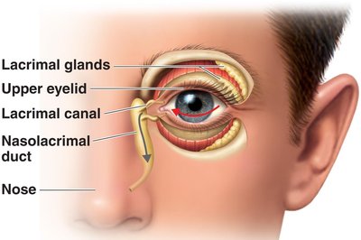
Lacrimal Apparatus and Other Washing Processes
The lacrimal apparatus protects the eyes by producing and draining tears, which wash away microbes. Other washing processes include salivation, urination, vaginal secretions, peristalsis, defecation, vomiting, and diarrhea, all of which help remove pathogens from the body.
Chemical Factors Affecting Microbes
Sebum: Oily substance from sebaceous glands that inhibits microbial growth.
Ear Wax: Physical barrier, high in fatty acids and sebum.
Lysozyme: Enzyme that breaks down peptidoglycan in bacterial cell walls; found in sweat, tears, saliva, nasal secretions, tissue fluids, and urine.
Lactic Acid: Lowers pH on skin and in vaginal secretions, inhibiting microbes.
Saliva: Contains uric acid (pH 6.55–6.85) and immunoglobulin A (IgA) antibodies.
Gastric Juice: Contains hydrochloric acid (pH 1.2–3), enzymes, and mucus.
Cervical Mucus: Some antimicrobial activity.
Urine: Contains urea and salts that inhibit microbial growth.
Normal Microbiota
Normal microbiota provide protection by competing with pathogens for nutrients and attachment sites, producing antimicrobial substances, and stimulating the immune system.
Competitive Exclusion: Outcompete pathogens for resources.
Microbial Antagonism: Produce antibiotics, alter environmental conditions (e.g., lower pH).
Immune System Development: Exposure to microbiota strengthens immune responses.
Probiotics: May enhance normal microbiota in unhealthy individuals.
Second Line of Defense
Blood Components
Blood consists of plasma and formed elements (cells and cell fragments). The main cellular components involved in innate immunity are leukocytes (white blood cells).
Erythrocytes: Red blood cells, transport oxygen and carbon dioxide.
Platelets: Cell fragments involved in blood clotting.
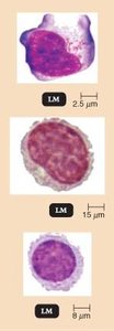
Leukocytes: White blood cells, divided into granulocytes and agranulocytes.
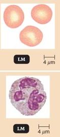
Neutrophils: Phagocytosis and diapedesis (movement into tissues).
Basophils: Produce histamine, involved in inflammation and allergic responses.
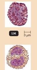
Eosinophils: Attack parasites, some phagocytosis, and diapedesis.
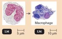
Monocytes: Mature into macrophages, perform phagocytosis.
Dendritic Cells: Phagocytosis and initiation of adaptive immune responses.
Lymphocytes: Include natural killer (NK) cells, T cells, and B cells.
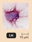
NK Cells: Destroy abnormal body cells by releasing perforin and granzymes.
T Cells: Cell-mediated immunity (third line of defense).
B Cells: Humoral immunity, produce antibodies (third line of defense).
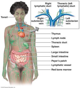
Lymphatic System
The lymphatic system is a network of vessels and organs that helps return interstitial fluid to the bloodstream and is involved in immune surveillance.
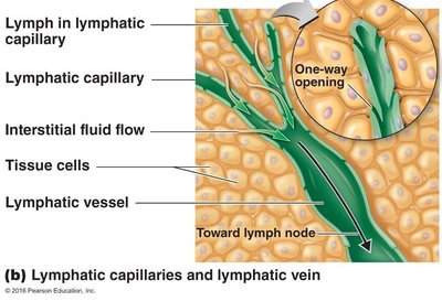
Lymph: Interstitial fluid that travels through lymphatic vessels.
Lymph Capillaries: Collect fluid from tissues.
Lymph Vessels: Transport lymph in one direction toward the heart.
Lymph Nodes: Filter lymph, trap microbes, and activate B and T cells.
Lymphatic Organs: Peyer’s patches (intestine), tonsils (throat), red bone marrow (origin of B and T cells), spleen (monitors blood), thymus (T cell maturation).
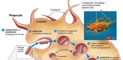
Phagocytosis
Phagocytosis is the process by which certain cells (neutrophils, eosinophils, dendritic cells, macrophages) ingest and destroy microbes and debris.
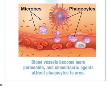
Chemotaxis: Phagocytes are attracted to infection sites by chemical signals.
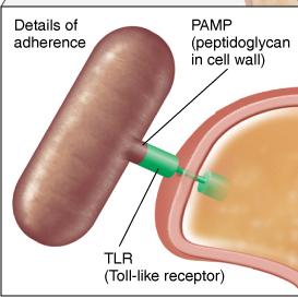
Adherence: Phagocyte receptors (TLRs) bind to pathogen-associated molecular patterns (PAMPs) on microbes.
Ingestion: Phagocyte engulfs the microbe, forming a phagosome.
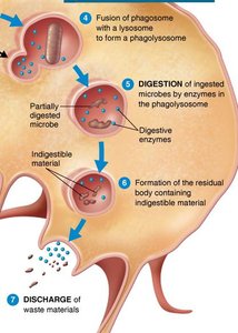
Phagolysosome Formation: Phagosome fuses with lysosome, forming a phagolysosome containing digestive enzymes.
Digestion: Microbes are broken down by enzymes.
Residual Body Formation: Indigestible material is contained in a residual body.
Discharge: Waste is expelled from the cell by exocytosis.
Microbial Evasion of Phagocytosis
Inhibit Adherence: Capsules prevent phagocyte binding.
Leukocidins: Toxins that destroy phagocytes.
Pore-forming Toxins: Cause phagocyte leakage.
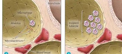
Avoid Digestion: Mycobacterium tuberculosis resists digestion due to mycolic acid.
Prevent Lysosome Fusion: HIV prevents phagolysosome formation.
Biofilms: Protect bacteria from phagocytosis.
Inflammation
Inflammation is a local defensive response to tissue injury or infection, characterized by pain, redness, immobility, swelling, and heat (PRISH).
Acute Inflammation: Short and intense.
Chronic Inflammation: Long-lasting, less intense, may cause tissue damage.
Blood Vessel Changes: Damaged cells release histamine, kinins, and cytokines, causing vasodilation and increased permeability.
Blood Clots: Prevent spread of microbes.
Abscess Formation: Local collection of pus.
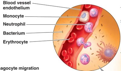
Phagocyte Migration: Margination (phagocytes stick to endothelium), diapedesis (phagocytes move into tissues), and phagocytosis of pathogens.
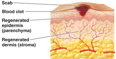
Tissue Repair: Parenchyma (functional tissue) regenerates; stroma (supporting tissue) may form scar tissue.
Fever
Fever is a systemic response to infection, raising body temperature above normal (37°C). High fevers above 44–46°C can be fatal. Fever helps inhibit microbial growth and enhances immune responses.
Complement System
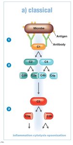
The complement system consists of plasma proteins that enhance immune responses through a cascade of activation. There are three main pathways:
Classical Pathway: Triggered by antigen-antibody complexes binding to C1, leading to activation of C2 and C4, and ultimately C3.
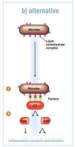
Alternative Pathway: Triggered by factors B, D, and P binding to microbial surfaces, leading to C3 activation.
Lectin Pathway: Triggered by lectins binding to mannose on pathogens, activating C2 and C4, and then C3.
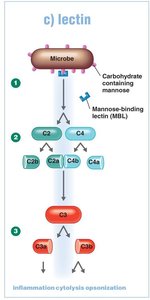
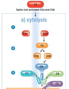
All pathways converge at C3, leading to:
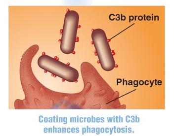
Opsonization: C3b coats microbes, enhancing phagocytosis.
Inflammation: C3a and C5a stimulate mast cells to release histamine, increasing blood vessel permeability.
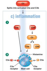
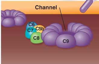
Cytolysis: C5b, C6, C7, C8, and C9 form the membrane attack complex (MAC), creating pores in microbial membranes and causing cell lysis.
Microbial Evasion of Complement: Capsules, altered LPS, and enzymes that degrade complement components help microbes avoid destruction.
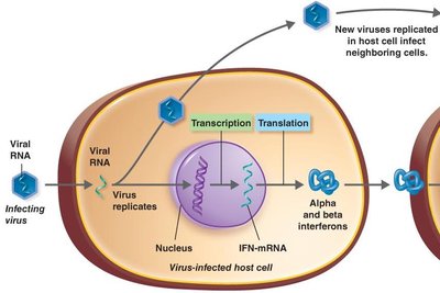
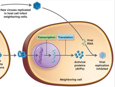
Interferons
Interferons are proteins produced by virus-infected cells that signal neighboring cells to produce antiviral proteins (AVPs), which inhibit viral replication.
Viral RNA enters host cell and replicates.
Infected cell produces and releases interferons.
Interferons bind to receptors on neighboring cells, inducing AVP production.
AVPs degrade viral mRNA and inhibit protein synthesis, preventing viral replication.
Antimicrobial Substances
Iron-binding Proteins: Transferrin, lactoferrin, ferritin, and hemoglobin bind iron, limiting its availability to microbes.
Antimicrobial Peptides: Dermcidin (sweat glands), defensins (produced by neutrophils) disrupt microbial membranes and inhibit growth.
