Back
BackInnate Immunity: The Body’s First and Second Lines of Defense
Study Guide - Smart Notes
 Tailored notes based on your materials, expanded with key definitions, examples, and context.
Infection, Infectious Diseases, and Epidemiology
Occurrence of Disease: Endemic, Epidemic, Pandemic, and Sporadic
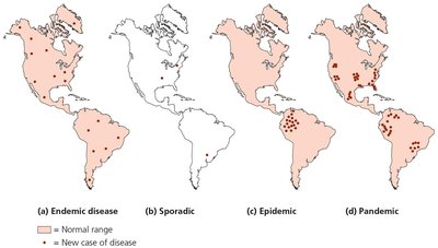
The occurrence of infectious diseases can be classified based on their frequency and geographic distribution. Understanding these terms is essential for epidemiology and public health.
Endemic Disease: A disease that is constantly present at a stable frequency within a particular geographic area.
Sporadic Disease: A disease that occurs infrequently and irregularly.
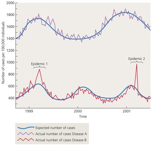
Epidemic Disease: A disease that occurs at a greater frequency than usual in a given area or population.
Pandemic Disease: An epidemic that occurs simultaneously on more than one continent.
Key Point: The number of cases alone does not determine whether a disease is an epidemic or pandemic; the expected baseline and geographic spread are critical factors.
Innate Immunity
Overview of the Body’s Defenses
Innate immunity refers to the nonspecific defense mechanisms that come into play immediately or within hours of an antigen's appearance in the body. These defenses are present at birth and provide the first and second lines of defense against pathogens.
Species Resistance: Many pathogens are unable to infect humans due to physiological incompatibilities, such as the absence of necessary receptors or unsuitable environmental conditions.
First Line of Defense: External physical barriers (skin and mucous membranes) and associated chemicals/processes.
Second Line of Defense: Internal defenses including protective cells, bloodborne chemicals, and processes that inactivate or kill invaders.
The Body’s First Line of Defense
The first line of defense consists of physical and chemical barriers that prevent pathogen entry.
The Role of Skin in Innate Immunity
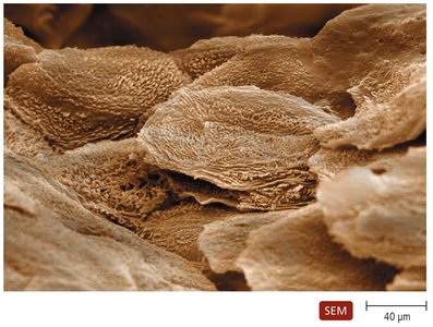
Epidermis: Multiple layers of tightly packed cells; shedding removes microorganisms; dendritic cells phagocytize pathogens.
Dermis: Collagen fibers provide strength and resistance to abrasions.
Chemical Defenses: Perspiration (contains salt, antimicrobial peptides, lysozyme), sebum (keeps skin pliable, lowers pH).
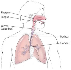
The Role of Mucous Membranes in Innate Immunity
Epithelium: Thin, living, tightly packed cells; continual shedding removes microbes; dendritic cells below epithelium phagocytize pathogens.
Goblet and Ciliated Columnar Cells: Produce mucus and move trapped pathogens out of the body.
Deeper Connective Layer: Supports the epithelium and produces defensive chemicals.
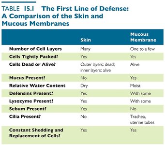
Comparison of Skin and Mucous Membranes
The following table compares the main features of skin and mucous membranes as barriers to infection:
Skin | Mucous Membrane | |
|---|---|---|
Number of Cell Layers | Many | One to a few |
Cells Tightly Packed? | Yes | Yes |
Cells Dead or Alive? | Outer layers: dead; inner layers: alive | Alive |
Mucus Present? | No | Yes |
Relative Water Content | Dry | Moist |
Defensins Present? | Yes | With some |
Lysozyme Present? | Yes | With some |
Sebum Present? | Yes | No |
Cilia Present? | No | Trachea, uterine tubes |
Constant Shedding and Replacement of Cells? | Yes | Yes |
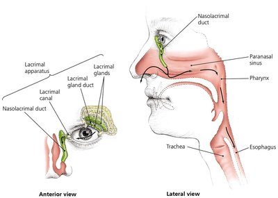
The Role of the Lacrimal Apparatus in Innate Immunity
Lacrimal Apparatus: Produces and drains tears; blinking spreads tears and washes the eye surface.
Lysozyme in Tears: Destroys bacterial cell walls.
The Role of the Microbiome in Innate Immunity
Microbial Antagonism (Competitive Inhibition): The microbiome competes with potential pathogens for nutrients and attachment sites.
Additional Benefits: Microbiome members create unfavorable environments for pathogens, stimulate the second line of defense, generate antimicrobial compounds, and provide vitamins to the host.
Other First-Line Defenses
Antimicrobial Peptides: Present in skin, mucous membranes, and neutrophils; act against a variety of microbes by disrupting membranes or interfering with metabolism.
Other Secretions and Activities: Many organs secrete chemicals with antimicrobial properties.
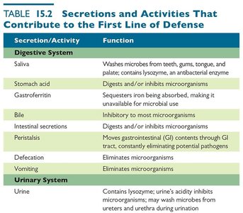
Secretions and Activities That Contribute to the First Line of Defense
Various body systems contribute to innate immunity through secretions and activities that inhibit or eliminate pathogens:
Secretion/Activity | Function |
|---|---|
Saliva | Washes microbes from teeth, gums, and palate; contains lysozyme |
Stomach acid | Digests and/or inhibits microorganisms |
Gastroferritin | Sequesters iron, making it unavailable for microbial use |
Bile | Inhibitory to most microorganisms |
Intestinal secretions | Digest and/or inhibit microorganisms |
Peristalsis | Moves GI contents, eliminating pathogens |
Defecation/Vomiting | Eliminates microorganisms |
Urine | Contains lysozyme; acidity inhibits microorganisms |
Vaginal secretions | Acidity inhibits microorganisms; contains iron-binding proteins |
Menstrual flow | Cleanses uterus and vagina |
Prostate secretion | Contains iron-binding proteins |
Blood flow | Removes microorganisms from wounds |
Coagulation | Prevents entrance of many pathogens |

The Body’s Second Line of Defense
The second line of defense is activated when pathogens penetrate the skin or mucous membranes. It includes cellular and chemical components found in the blood and tissues.
Defense Components of Blood
Plasma: Contains water, electrolytes, dissolved gases, nutrients, proteins (including complement proteins and antibodies), and iron-binding compounds.
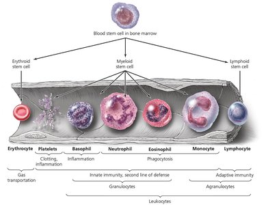
Formed Elements: Erythrocytes (transport gases), platelets (clotting), and leukocytes (defense against invaders).
Leukocytes: Divided into granulocytes (basophils, eosinophils, neutrophils) and agranulocytes (lymphocytes, monocytes).
Lab Analysis of Leukocytes
Differential White Blood Cell Count: Used to diagnose infections and diseases.
Increased Eosinophils: Indicate allergies or parasitic worm infection.
Bacterial Infections: Often show increased leukocytes and neutrophils.
Viral Infections: Often show increased lymphocytes.
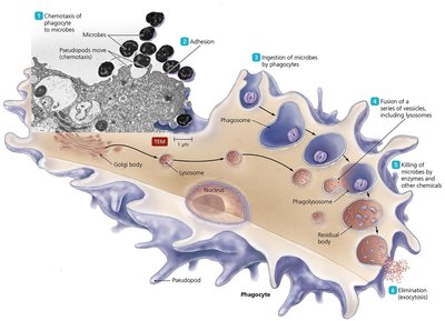
Phagocytosis
Phagocytosis is the process by which certain cells (phagocytes) ingest and destroy pathogens. It involves six stages:
Chemotaxis
Adhesion
Ingestion
Maturation
Killing
Elimination
Nonphagocytic Killing
Eosinophils: Attack parasitic helminths by secreting toxins.
Natural Killer (NK) Cells: Secrete toxins onto the surface of virally infected cells and tumors.
Neutrophils: Produce chemicals and extracellular traps (NETs) to kill bacteria.
Nonspecific Chemical Defenses Against Pathogens
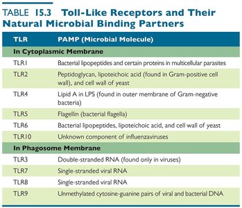
Toll-like Receptors (TLRs): Integral membrane proteins on phagocytes that recognize pathogen-associated molecular patterns (PAMPs) and initiate defensive responses such as inflammation, apoptosis, and stimulation of adaptive immunity.
NOD Proteins: Cytosolic proteins that bind PAMPs and trigger inflammation and other innate responses.
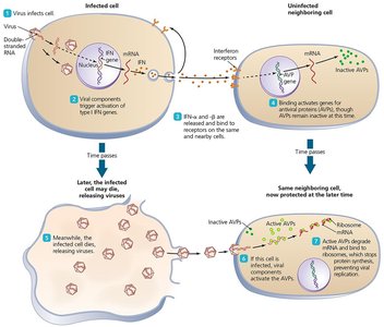
Interferons: Proteins released by host cells to inhibit the spread of viral infections. Two types: Type I (alpha and beta) and Type II (gamma).
TLR | PAMP (Microbial Molecule) |
|---|---|
TLR1 | Bacterial lipopeptides and certain proteins in multicellular parasites |
TLR2 | Peptidoglycan, lipoteichoic acid (Gram-positive cell wall), and cell wall of yeast |
TLR4 | Lipid A in LPS (Gram-negative bacteria) |
TLR5 | Flagellin (bacterial flagella) |
TLR6 | Bacterial lipopeptides, lipoteichoic acid, and cell wall of yeast |
TLR10 | Unknown component of influenzaviruses |
TLR3 | Double-stranded RNA (viruses) |
TLR7 | Single-stranded viral RNA |
TLR8 | Single-stranded viral RNA |
TLR9 | Unmethylated cytosine-guanine pairs of viral and bacterial DNA |
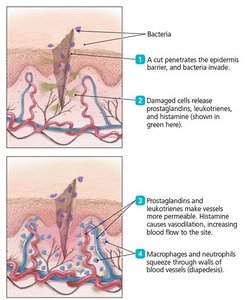
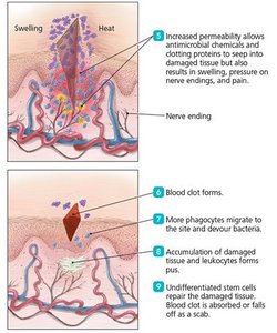
Inflammation
Inflammation is a nonspecific response to tissue damage, characterized by redness, heat, swelling, and pain. It can be acute (short-lived and beneficial) or chronic (long-lasting and potentially damaging).
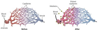
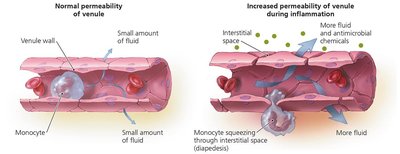
Acute Inflammation: Involves vasodilation, increased permeability of blood vessels, migration of phagocytes, and tissue repair.
Chronic Inflammation: Can result in tissue damage and disease.
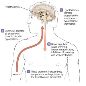
Fever
Fever is a systemic response to infection, defined as a body temperature above 37°C. It is triggered by pyrogens, which reset the hypothalamic thermostat. Fever enhances the effects of interferons, inhibits the growth of some microbes, and may enhance the activities of phagocytes and tissue repair.
Pyrogens: Include bacterial toxins, cytoplasmic contents of bacteria, antibody-antigen complexes, and substances released by phagocytes.
Mechanism: Not fully understood, but involves the hypothalamus and various immune mediators.