Back
Innate Immunity: The Body’s First and Second Lines of Defense
Study Guide - Smart Notes
 Tailored notes based on your materials, expanded with key definitions, examples, and context.
Innate Immunity
Overview of Host Defenses
Innate immunity is the body’s first response to invading pathogens and is present from birth. It provides immediate, nonspecific defense and does not generate immunological memory. In contrast, adaptive immunity develops over time and is characterized by specificity and memory.
Species Resistance: Human cells are naturally incompatible with most pathogens, providing a basic level of protection.
Innate Defense: Always present and ready to act against invaders.
Adaptive Defense: Develops after exposure to specific pathogens and involves immunological memory.
The Body’s First Line of Defense
Physical and Chemical Barriers
The first line of defense consists of structures, chemicals, and processes that prevent pathogens from entering the body. The primary components are the skin and mucous membranes, which line the respiratory, digestive, urinary, and reproductive systems.
Skin: Acts as a physical barrier with tightly packed cells and antimicrobial chemicals.
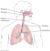
Mucous Membranes: Line all body cavities open to the environment and trap pathogens.
The Role of Skin in Innate Immunity
The skin is composed of two main layers:
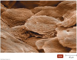
Epidermis: Multiple layers of tightly packed cells prevent pathogen entry. Shedding of dead cells removes microorganisms. Epidermal dendritic cells phagocytize pathogens.
Dermis: Contains collagen fibers that resist abrasions, reducing the risk of pathogen entry.
Chemical Defenses: Perspiration (sweat) contains salt and antimicrobial peptides; lysozyme destroys bacterial cell walls. Sebum (oil) keeps skin pliable and lowers pH, inhibiting bacterial growth.
The Role of Mucous Membranes in Innate Immunity
Mucous membranes consist of two layers:
Epithelium: Tightly packed cells prevent pathogen entry. Goblet and ciliated columnar cells help remove invaders.
Deeper Connective Layer: Supports the epithelium.
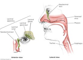
The Role of the Lacrimal Apparatus in Innate Immunity
The lacrimal apparatus produces and drains tears, which contain lysozyme that destroys bacteria, protecting the eyes from infection.
The Role of the Microbiome in Innate Immunity
The normal microbiome provides protection through microbial antagonism, making it difficult for pathogens to colonize the body by:
Consuming nutrients
Creating an unfavorable environment for other microbes
Preventing pathogen attachment to host cells
Producing antimicrobial compounds
Other First-Line Defenses
Antimicrobial Peptides: Present in skin, mucous membranes, and neutrophils; act against a variety of microbes through multiple mechanisms.
Other Chemicals: Many organs secrete chemicals with antimicrobial properties.
The Body’s Second Line of Defense
General Features
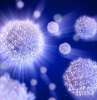
The second line of defense is activated when pathogens penetrate the skin or mucous membranes. It includes cells, antimicrobial chemicals, and processes, many of which are found in or originate from the blood.
Defense Components of Blood
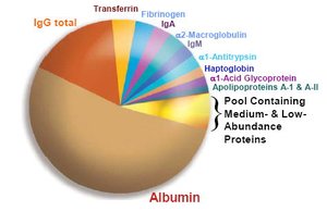
Plasma: The liquid portion of blood, mostly water and dissolved proteins, including complement proteins and antibodies.
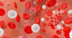
Leukocytes (White Blood Cells): Involved in defending the body against invaders. Divided into granulocytes and agranulocytes.
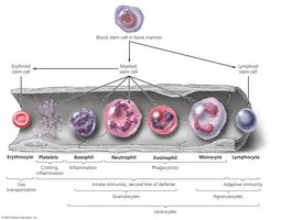
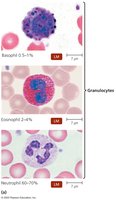
Granulocytes
Basophils: Stain blue with basic dye; release inflammatory chemicals but do not phagocytize.
Eosinophils: Stain red/orange with acidic dye; phagocytize pathogens and are capable of diapedesis.
Neutrophils: Stain lilac with a mix of dyes; phagocytize pathogens and are capable of diapedesis.
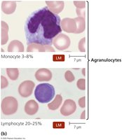
Agranulocytes
Lymphocytes: Most involved in adaptive immunity; natural killer lymphocytes are part of innate immunity.
Monocytes: Leave the blood and mature into macrophages, which are phagocytic cells that devour foreign objects.
Lab Analysis of Leukocytes
Differential white blood cell count can indicate disease.
Increased eosinophils suggest allergies or parasitic worm infection.
Bacterial diseases often show increased leukocytes and neutrophils.
Viral infections show increased lymphocytes.
Phagocytosis
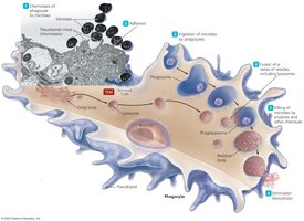
Phagocytosis is the process by which certain cells (phagocytes) ingest and destroy pathogens. It occurs in six stages:
Chemotaxis
Adhesion
Ingestion
Maturation
Killing
Elimination
Nonphagocytic Killing
Eosinophils and Natural Killer (NK) Cells: Attack pathogens with toxins.
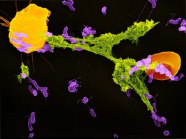
Neutrophils: Generate extracellular fibers called neutrophil extracellular traps (NETs) that bind and kill bacteria.
Nonspecific Chemical Defenses Against Pathogens
Toll-like Receptors (TLRs): Bind pathogen-associated molecular patterns (PAMPs) and initiate defensive responses, including secretion of inflammatory mediators, stimulation of adaptive immunity, and apoptosis.
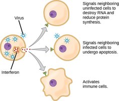
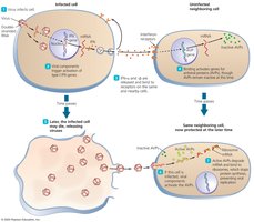
Interferons: Protein molecules released by host cells to inhibit the spread of viral infections nonspecifically.
Complement System
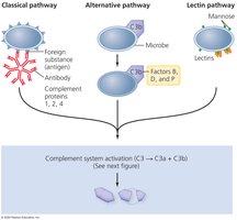
The complement system is a set of serum proteins that, when activated, result in the lysis of foreign cells and trigger inflammation and fever. Complement proteins are designated numerically according to their order of discovery.
Inflammation
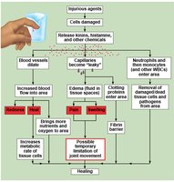
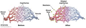
Inflammation is characterized by redness, heat, swelling, and pain. Acute inflammation develops quickly, is short-lived, and is typically beneficial. It is important in the second line of defense and involves:
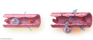
Dilation and increased permeability of blood vessels
Migration of phagocytes to the site of infection
Tissue repair
Migration of Phagocytes and Tissue Repair
Neutrophils and monocytes are delivered to the site of infection, recruited by chemotactic factors, and enter the site by squeezing between vessel wall cells (diapedesis).
Delivery of nutrients and oxygen to the site facilitates tissue repair.
Fever
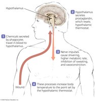
Fever is defined as a body temperature above 37°C and results when pyrogens trigger the hypothalamus to increase the body’s core temperature. Pyrogens include bacterial toxins, cytoplasmic contents of lysed bacteria, antibody-antigen complexes, and substances released by phagocytes after ingesting bacteria.
Fever continues as long as pyrogens are present.
Outcomes of fever include enhanced effects of interferons, inhibition of some microbial growth, and enhanced activity of phagocytes, immune cells, and tissue repair processes.