 Back
BackInnate Immunity: The Body’s First and Second Lines of Defense
Study Guide - Smart Notes
 Tailored notes based on your materials, expanded with key definitions, examples, and context.
Innate Immunity
Overview of the Body’s Defenses
Innate immunity provides the body’s initial defense against pathogens through physical, chemical, and cellular mechanisms. It is non-specific and acts rapidly to prevent infection and disease.
Species resistance: Humans are naturally resistant to many pathogens due to physiological incompatibilities, such as the absence of necessary receptors or unsuitable environmental conditions for the pathogen.
Limitations: Despite these defenses, humans are still susceptible to a range of pathogens.
The Body’s First Line of Defense
Physical and Chemical Barriers
The first line of defense consists of structures, chemicals, and processes that prevent pathogens from entering the body. The primary barriers are the skin and mucous membranes.
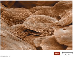
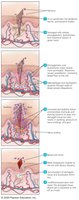
Skin: Acts as a physical barrier with tightly packed cells and continual shedding, which removes microorganisms. The skin also contains dendritic cells that phagocytize pathogens.
Mucous membranes: Line all body cavities open to the environment, consisting of a living epithelium and a supportive connective tissue layer. These membranes trap and remove invaders through mucus and ciliary action.
Chemical Defenses of the Skin
Perspiration: Contains salt (inhibits microbial growth), antimicrobial peptides, and lysozyme (destroys bacterial cell walls).
Sebum: Secreted by sebaceous glands, keeps skin pliable and lowers pH, inhibiting bacterial growth.
The Role of Mucous Membranes in Innate Immunity
Epithelium: Thin, living, tightly packed cells that are continually shed, removing microbes.
Dendritic cells: Located below the epithelium, these cells phagocytize pathogens.
Goblet and ciliated columnar cells: Produce mucus and move trapped particles out of the body.
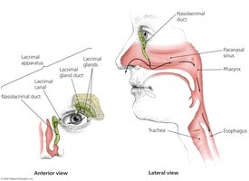
Lacrimal Apparatus
The lacrimal apparatus produces and drains tears, which contain lysozyme to destroy bacteria and help wash the surface of the eye.
The Role of the Microbiome in Innate Immunity
Microbial antagonism: The normal microbiome competes with pathogens for nutrients and attachment sites, produces antimicrobial compounds, and stimulates the immune system.
Health benefits: The microbiome also provides vitamins and modulates immune responses.
Other First-Line Defenses
Antimicrobial peptides: Present in skin, mucous membranes, and neutrophils, these peptides disrupt microbial membranes and interfere with pathogen metabolism.
Secretions: Many organs secrete chemicals with antimicrobial properties, contributing to the first line of defense.
The Body’s Second Line of Defense
Defense Components of Blood
When pathogens breach the first line of defense, the second line is activated, involving cells, antimicrobial chemicals, and processes found in the blood.
Plasma: The liquid portion of blood containing electrolytes, nutrients, proteins (including complement proteins and antibodies), and iron-binding compounds.
Formed elements: Includes erythrocytes (oxygen and CO2 transport), platelets (clotting), and leukocytes (defense against invaders).
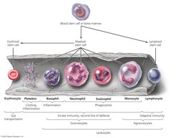
Leukocytes: Granulocytes and Agranulocytes
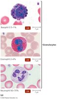
Granulocytes: Contain granules that stain differently. Types include:
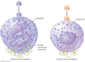
Basophils: Release inflammatory chemicals.
Eosinophils: Phagocytize pathogens and are involved in defense against parasites and allergies.
Neutrophils: Phagocytize pathogens and are the most abundant leukocytes.
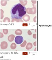
Agranulocytes: Cytoplasm appears uniform. Types include:
Lymphocytes: Key cells in adaptive immunity and natural killer (NK) cells.
Monocytes: Mature into macrophages, which are phagocytic cells.
Lab Analysis of Leukocytes
Differential white blood cell count: Used to diagnose infections and diseases.
Increased eosinophils: Allergies or parasitic infections.
Increased neutrophils: Bacterial infections.
Increased lymphocytes: Viral infections.
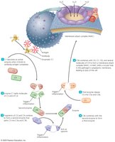
Phagocytosis
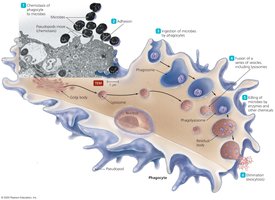
Phagocytosis is the process by which certain cells (phagocytes) ingest and destroy pathogens. It occurs in six stages:
Chemotaxis
Adhesion
Ingestion
Maturation
Killing
Elimination
Nonphagocytic Killing
Eosinophils: Attack parasitic helminths by secreting toxins and can form extracellular structures to kill bacteria.
Natural killer (NK) cells: Secrete toxins onto virally infected cells and tumors, distinguishing them from normal cells.
Neutrophils: Produce chemicals and extracellular traps (NETs) to kill microbes without phagocytosis.
Nonspecific Chemical Defenses Against Pathogens
Toll-like Receptors (TLRs) and NOD Proteins
TLRs: Integral membrane proteins on phagocytes that recognize pathogen-associated molecular patterns (PAMPs) and trigger defensive responses, including inflammation and apoptosis.
NOD proteins: Cytosolic proteins that bind PAMPs and trigger innate immune responses.
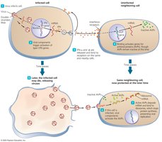
Interferons
Interferons are proteins released by host cells to inhibit the spread of viral infections. They induce antiviral states in neighboring cells and are responsible for many symptoms of viral infections.
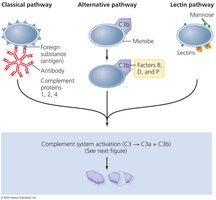
Complement System
The complement system is a group of serum proteins that, when activated, lead to the lysis of foreign cells, inflammation, and enhanced phagocytosis. There are three activation pathways:
Classical pathway
Alternative pathway
Lectin pathway
Inflammation
General Features of Inflammation
Inflammation is a nonspecific response to tissue damage, characterized by redness, heat, swelling, and pain. It can be acute (short-lived and beneficial) or chronic (long-lasting and potentially damaging).
Acute inflammation: Involves vasodilation, increased vascular permeability, migration of phagocytes, and tissue repair.
Chronic inflammation: Can result in tissue damage and disease.
Inflammatory Mediators
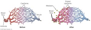
Vasodilation: Increases blood flow, causing redness and heat.
Chemicals involved: Bradykinins, prostaglandins, leukotrienes, and histamine.
Blood clotting proteins: Delivered to the site of injury to prevent pathogen spread.
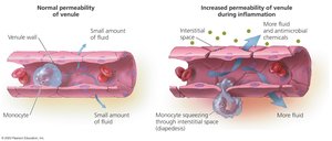
Mechanism of Inflammation
Migration of phagocytes: Neutrophils and monocytes are recruited to the site of infection, attach to blood vessel receptors, and migrate into tissues (diapedesis).
Tissue repair: Enhanced by increased delivery of nutrients and oxygen, though some tissues may not fully recover.
Fever
What is Fever?
Fever is an elevation of body temperature above 37°C, triggered by pyrogens that act on the hypothalamus. Pyrogens include bacterial toxins, cytoplasmic contents of lysed bacteria, antibody-antigen complexes, and substances released by phagocytes.
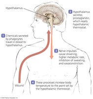
Why Fever Occurs
Fever persists as long as pyrogens are present.
Benefits of fever:
Enhances the effects of interferons.
Inhibits growth of some microbes.
May enhance phagocyte activity, adaptive immunity, and tissue repair.
Summary Table: Nonspecific Components of Innate Immunity
First Line of Defense | Second Line of Defense |
|---|---|
Skin, mucous membranes, microbiome, antimicrobial peptides, secretions (e.g., tears, saliva, stomach acid) | Phagocytes, inflammation, fever, complement system, interferons, antimicrobial peptides |
