 Back
BackInnate Immunity: The Body’s First and Second Lines of Defense
Study Guide - Smart Notes
 Tailored notes based on your materials, expanded with key definitions, examples, and context.
Innate Immunity: An Overview
Introduction to Innate Immunity
Innate immunity refers to the nonspecific defense mechanisms that come into play immediately or within hours of an antigen's appearance in the body. These defenses are present from birth and provide the first and second lines of defense against pathogens, acting before adaptive immunity is activated.
Species Resistance: Humans are naturally resistant to many pathogens that affect other species due to physiological and biochemical incompatibilities.
First Line of Defense: Physical and chemical barriers such as skin, mucous membranes, and associated secretions.
Second Line of Defense: Cellular and chemical responses that act when pathogens breach the first line, including phagocytic cells, inflammation, fever, and antimicrobial proteins.
The Body’s First Line of Defense
Physical Barriers: Skin and Mucous Membranes
The skin and mucous membranes form the primary physical barriers to pathogen entry. These structures are supported by chemical defenses that inhibit microbial growth and facilitate removal of invaders.
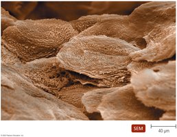
Skin: Composed of the epidermis (multiple layers of tightly packed cells) and dermis (collagen fibers for strength). The shedding of dead skin cells removes attached microorganisms, and dendritic cells in the epidermis phagocytize pathogens.
Chemical Defenses of Skin:
Perspiration (sweat) contains salt, antimicrobial peptides, and lysozyme, which destroys bacterial cell walls.
Sebum (oil) keeps skin pliable and lowers pH, inhibiting bacterial growth.
Mucous Membranes: Line all body cavities open to the environment. The epithelium is a thin, living layer of tightly packed cells that are continually shed, removing microbes. Goblet cells produce mucus, and ciliated cells help move trapped particles out. Dendritic cells below the epithelium phagocytize invaders.
Other First-Line Defenses
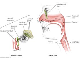
Lacrimal Apparatus: Produces and drains tears, which contain lysozyme to destroy bacteria and physically wash the eye surface.
Normal Microbiome: Competes with pathogens for nutrients and attachment sites, produces antimicrobial compounds, and stimulates the immune system. This microbial antagonism is crucial for preventing colonization by pathogens.
Antimicrobial Peptides: Found in skin, mucous membranes, and neutrophils; these peptides disrupt microbial membranes and interfere with pathogen metabolism.
Other Secretions: Many organs secrete chemicals with antimicrobial properties, such as stomach acid, bile, and urine.
The Body’s Second Line of Defense
Defense Components of Blood
When pathogens penetrate the first line of defense, the second line is activated. This includes cellular and chemical components found in blood.
Plasma: The liquid portion of blood containing water, electrolytes, nutrients, proteins (including complement proteins and antibodies), and iron-binding compounds.
Formed Elements:
Erythrocytes (RBCs): Transport oxygen and carbon dioxide.
Platelets: Involved in blood clotting.
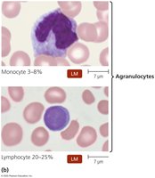
Leukocytes (WBCs): Defend against invaders and are divided into granulocytes and agranulocytes.
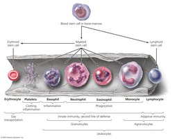
Leukocytes: Granulocytes and Agranulocytes
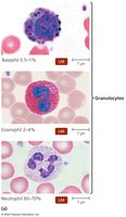
Granulocytes: Contain visible granules in their cytoplasm.
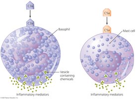
Basophils: Release inflammatory chemicals; stain blue with basic dye.
Eosinophils: Phagocytize pathogens and attack parasitic worms; stain red/orange with acidic dye.
Neutrophils: Phagocytize pathogens and are capable of diapedesis; stain lilac with a mix of dyes.
Agranulocytes: Lack visible granules.
Lymphocytes: Most involved in adaptive immunity; include natural killer (NK) cells.
Monocytes: Leave blood to mature into macrophages, which are phagocytic.
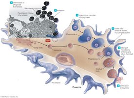
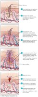
Phagocytosis
Phagocytosis is a multi-step process by which certain cells (phagocytes) ingest and destroy pathogens. The process includes:
Chemotaxis: Movement toward chemical signals from pathogens or damaged tissue.
Adhesion: Attachment of phagocyte to microbe.
Ingestion: Engulfment of the microbe into a phagosome.
Maturation: Fusion of phagosome with lysosome to form phagolysosome.
Killing: Destruction of the microbe by enzymes and toxic substances.
Elimination: Expulsion of debris from the cell.
Nonphagocytic Killing Mechanisms
Eosinophils: Attack parasitic worms by secreting toxins and can release mitochondrial DNA to trap and kill bacteria.
Natural Killer (NK) Cells: Secrete toxins onto the surface of virally infected cells and tumors, inducing apoptosis.
Neutrophils: Can release chemicals and form neutrophil extracellular traps (NETs) to bind and kill bacteria without phagocytosis.
Nonspecific Chemical Defenses
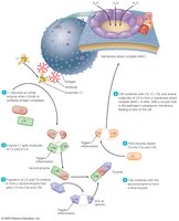
Toll-like Receptors (TLRs): Integral membrane proteins on phagocytes that recognize pathogen-associated molecular patterns (PAMPs) and trigger immune responses such as inflammation and apoptosis.
NOD Proteins: Cytosolic proteins that detect PAMPs inside the cell and initiate inflammation and other responses.
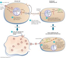
Interferons (IFNs): Proteins released by host cells in response to viral infection; they inhibit viral replication and activate immune cells.
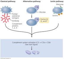
Complement System: A group of serum proteins that, when activated, lead to lysis of pathogens, inflammation, and enhanced phagocytosis. Activation occurs via three pathways: classical, alternative, and lectin.
Inflammation
General Features of Inflammation
Inflammation is a nonspecific response to tissue damage caused by infection or injury. It is characterized by redness, heat, swelling, and pain. There are two types:
Acute Inflammation: Rapid onset, short duration, and typically beneficial for eliminating pathogens and repairing tissue.
Chronic Inflammation: Long-lasting and can result in tissue damage and disease.
Mechanisms and Mediators of Inflammation
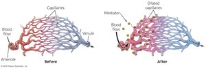
Vasodilation: Increases blood flow, causing redness and heat. Mediated by chemicals such as bradykinins, prostaglandins, leukotrienes, and histamine.
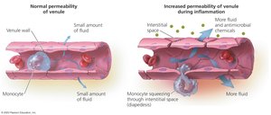
Increased Vascular Permeability: Allows immune cells and proteins to enter tissues, leading to swelling.
Migration of Phagocytes: Neutrophils and monocytes move to the site of infection, attracted by chemotactic factors.
Tissue Repair: Enhanced by increased delivery of nutrients and oxygen; some tissues may not fully recover.
Fever
Definition and Mechanism
Fever is an elevation of body temperature above 37°C, triggered by pyrogens that act on the hypothalamus. Pyrogens include bacterial toxins, cytoplasmic contents of lysed bacteria, antibody-antigen complexes, and substances released by phagocytes.
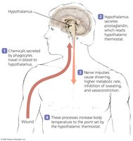
Benefits of Fever:
Enhances the effects of interferons
Inhibits growth of some microbes
May enhance phagocyte activity, adaptive immunity, and tissue repair
Summary Table: Nonspecific Components of Innate Immunity
First Line of Defense | Second Line of Defense |
|---|---|
Skin, mucous membranes, secretions, normal microbiome, antimicrobial peptides | Phagocytes, inflammation, fever, complement, interferons, NK cells, antimicrobial proteins |
