 Back
BackInnate Immunity: The Body’s First Line of Defense
Study Guide - Smart Notes
 Tailored notes based on your materials, expanded with key definitions, examples, and context.
Chapter 16: Innate Immunity
Overview of Innate vs. Adaptive Immunity
Immunity is the body's ability to resist infection and disease. It is divided into two main types: innate immunity and adaptive immunity. Innate immunity acts as the first responder, r'''ecognizing pathogens through predetermined molecular patterns, while adaptive immunity responds later, learning and remembering specific pathogens for future defense.
Innate Immunity: Immediate response, recognizes common pathogen-associated molecular patterns (PAMPs) via Toll-like receptors (TLRs), no memory.
Adaptive Immunity: Delayed response, highly specific, develops memory after exposure to pathogens.
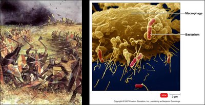
Image explanation: The left panel uses a battlefield analogy to represent the innate immune system's rapid, non-specific response. The right panel shows a macrophage (yellow) engulfing bacteria (red), illustrating phagocytosis, a key innate defense mechanism.
First Line of Defense: Physical and Chemical Barriers
Physical Barriers
The first line of defense prevents microbial entry through physical structures and actions:
Skin: Composed of keratinized, tightly packed epithelial cells, forming a tough barrier.
Mucous Membranes: Line the gastrointestinal, genitourinary, and respiratory tracts; secrete mucus to trap microbes.
Ciliary Escalator: Cilia in the respiratory tract move trapped particles upward, away from the lungs.
Other Physical Factors: Saliva, urine, vaginal secretions, and the lacrimal apparatus (tears) wash away microbes.
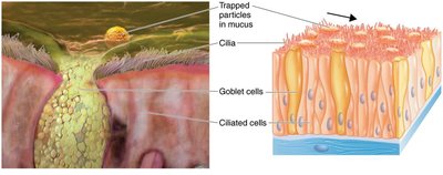
Image explanation: The left panel shows particles trapped in mucus, while the right panel details the structure of ciliated and goblet cells in the mucous membrane, illustrating the ciliary escalator mechanism.
Chemical Barriers
Chemical factors enhance the effectiveness of physical barriers:
Sebum: Oily secretion from sebaceous glands inhibits microbial growth.
Gastric Juices: Highly acidic environment in the stomach destroys most pathogens.
Vaginal Secretions: Acidic pH inhibits microbial colonization.
Role of Normal Flora
Normal flora (resident microbiota) compete with pathogens for nutrients and attachment sites, a process called microbial antagonism or competitive exclusion.
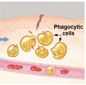
Second Line of Defense: Cellular and Molecular Components
Cells of the Innate Immune System
When pathogens breach the first line of defense, the second line involves various blood cells:
Red Blood Cells (Erythrocytes): Carry oxygen (not directly involved in immunity).
White Blood Cells (Leukocytes): Key players in immune defense, divided into granulocyte s and agranulocytes.
Granulocytes
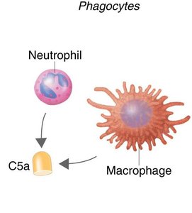
Neutrophils: First responders, phagocytic, most abundant (60–70%).
Basophils: Release histamine, involved in inflammation and allergic responses.
Eosinophils: Combat parasitic worms, involved in allergic reactions.
Agranulocytes
Monocytes: Circulate in blood, mature into macrophages in tissues, phagocytic.
Dendritic Cells: Phagocytic, bridge innate and adaptive immunity by presenting antigens.
Lymphocytes: Include T cells (cell-mediated immunity), B cells (antibody production), and Natural Killer (NK) cells (nonspecific killing of infected cells).
Differential White Cell Count
Cell Type | Percentage (%) |
|---|---|
Neutrophils | 60–70 |
Lymphocytes | 20–25 |
Monocytes | 3–8 |
Eosinophils | 2–4 |
Basophils | 0.5–1 |
Mnemonic: Never Let Monkeys Eat Bananas (order of abundance).
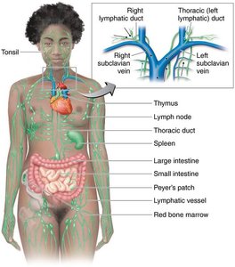
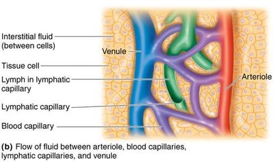
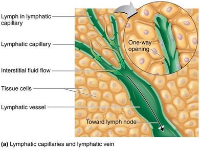
Lymphatic System
The lymphatic system is essential for immune cell circulation and pathogen clearance. It includes lymphatic vessels, primary lymphoid organs (bone marrow, thymus), and secondary lymphoid organs (lymph nodes, spleen, tonsils, Peyer's patches).
-
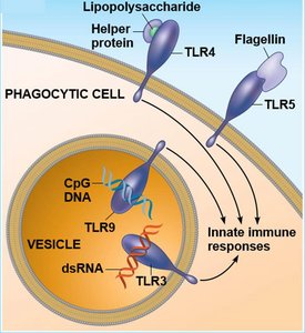
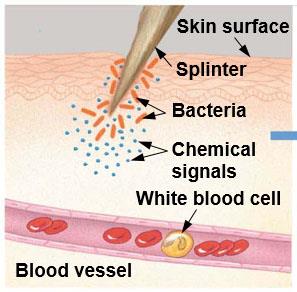
Recognition of Pathogens: Pattern Recognition Receptors
Innate immune cells recognize pathogens using Toll-like receptors (TLRs) that bind to pathogen-associated molecular patterns (PAMPs) such as lipopolysaccharide, flagellin, and unmethylated CpG DNA.
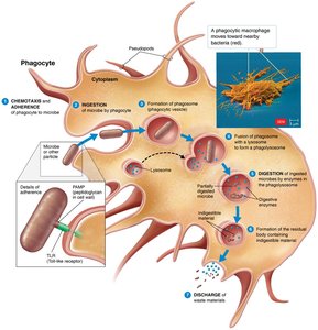
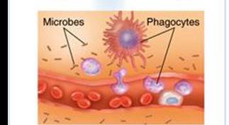
Phagocytosis: Mechanism and Evasion
Phases of Phagocytosis
Phagocytosis is the process by which cells (e.g., neutrophils, macrophages) ingest and destroy microbes:
Chemotaxis and Adherence: Phagocyte moves toward and attaches to microbe.
Ingestion: Microbe is engulfed into a phagosome.
Digestion: Phagosome fuses with lysosome, enzymes digest microbe.
Discharge: Indigestible material is expelled.

Microbial Evasion of Phagocytosis
Some microbes evade phagocytosis by producing capsules, protein A, protein M, or mycolic acid, which prevent attachment or digestion. Others produce leukocidins that lyse phagocytes.
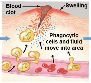
Inflammation
Inflammation is a localized response to infection or injury, characterized by redness, warmth, swelling, and pain. It has three main stages:
Vasodilation and Increased Permeability: Blood vessels widen, allowing immune cells to access the site.
Phagocyte Migration and Phagocytosis: Neutrophils and monocytes migrate to the site and ingest microbes.
Tissue Repair: Damaged tissue is repaired after the threat is eliminated.
Fever
Fever is an increase in body temperature above normal, often caused by infection. It inhibits microbial growth and enhances the activity of immune cells such as macrophages.
Antimicrobial Substances
Several substances in blood and tissues help eliminate microbes:
Complement System: A group of 30 proteins that, when activated, enhance phagocytosis, inflammation, and cytolysis.
Other Substances: Nitric oxide, superoxide, hydrogen peroxide, defensins, transferrins, and interferons.
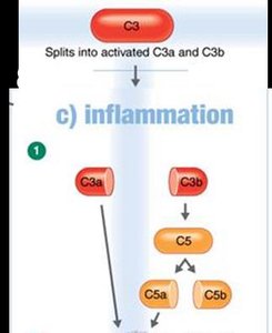
The Complement System
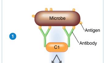
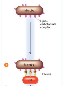
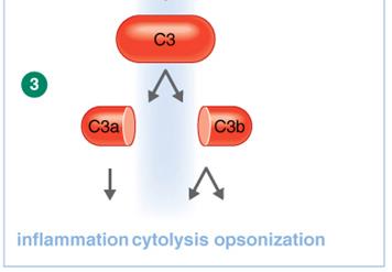
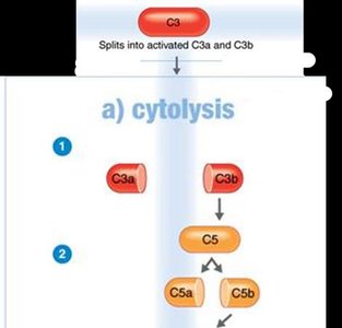
The complement system is activated via three pathways, all leading to the cleavage of C3 and subsequent immune responses:
Classical Pathway: Triggered by antigen-antibody complexes.
Alternative Pathway: Triggered by microbial surfaces.
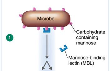
Lectin Pathway: Triggered by mannose-binding lectin binding to microbial carbohydrates.
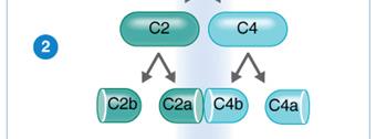
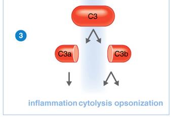
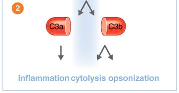
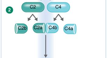
Outcomes of Complement Activation
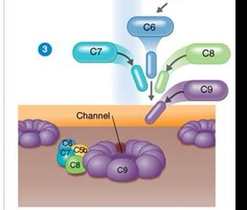
Cytolysis: Formation of the membrane attack complex (MAC) that creates pores in microbial membranes, leading to cell lysis.
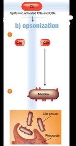
Opsonization: Coating of microbes with C3b, enhancing phagocytosis.
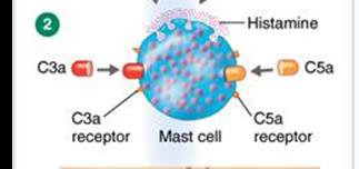
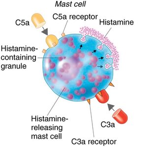
Inflammation: C3a and C5a stimulate mast cells to release histamine, increasing vascular permeability and attracting phagocytes.
Interferons and Cytokines
Interferons (IFNs) are cytokines that provide nonspecific defense against viruses:
IFN-γ: Activates macrophages.
IFN-α and IFN-β: Produced by virus-infected cells; induce neighboring cells to produce antiviral proteins (AVPs) that inhibit viral replication.
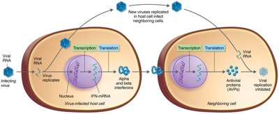
Image explanation: Virus-infected cells release interferons, which signal neighboring cells to produce AVPs, blocking viral replication.
Summary Table: Major Components of Innate Immunity
Component | Function | Example |
|---|---|---|
Physical Barriers | Prevent entry of microbes | Skin, mucous membranes |
Chemical Barriers | Destroy or inhibit microbes | Gastric acid, lysozyme |
Phagocytes | Ingest and destroy microbes | Neutrophils, macrophages |
Inflammation | Recruit immune cells, contain infection | Histamine release, vasodilation |
Fever | Inhibit microbial growth, enhance immunity | Increased body temperature |
Complement System | Opsonization, cytolysis, inflammation | C3, C5, MAC |
Interferons | Antiviral defense | IFN-α, IFN-β, IFN-γ |
Additional info: Some details, such as the mnemonic for white cell counts and the specific mechanisms of microbial evasion, were expanded for clarity and completeness.
