 Back
BackInnate Immunity: The First Line of Defense in Microbiology
Study Guide - Smart Notes
 Tailored notes based on your materials, expanded with key definitions, examples, and context.
Innate Immunity: Overview
Introduction to Innate Immunity
Innate immunity is the body's immediate, nonspecific defense mechanism against a wide variety of pathogens, including bacteria, viruses, fungi, protozoa, and parasitic worms. Unlike adaptive immunity, innate immunity is present from birth, acts rapidly, and does not develop immunological memory. It serves as the foundation for the immune system, working in concert with adaptive immunity for comprehensive protection.
Nonspecific: Targets a broad range of invaders, not specific pathogens.
Innate: Built-in and always active, not learned or improved over time.
Rapid Response: Acts immediately or within hours of pathogen exposure.
Limitations: Some pathogens can evade or overcome innate defenses; responses may sometimes damage host tissues.
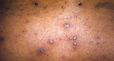
Example: The inflammation seen in chickenpox is a result of the innate immune response to viral infection.
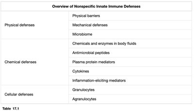
Categories of Nonspecific Innate Immunity
Major Components
Innate immunity consists of three overlapping categories that function together to prevent infection:
Physical defenses
Chemical defenses
Cellular defenses
Overview of Nonspecific Innate Immune Defenses | |
|---|---|
Physical defenses | Physical barriers, Mechanical defenses, Microbiome |
Chemical defenses | Chemicals and enzymes in body fluids, Antimicrobial peptides, Plasma protein mediators, Cytokines, Inflammation-eliciting mediators |
Cellular defenses | Granulocytes, Agranulocytes |

Physical Defenses
Physical Barriers
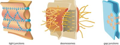
Physical barriers are the body's first line of defense, preventing microbes from reaching susceptible tissues. These barriers are formed by tightly joined cells, including epithelial and endothelial cells, which are reinforced by specialized cell junctions.
Tight junctions: Seal adjacent cells, preventing passage of pathogens.
Desmosomes: Provide structural support, allowing limited material passage.
Gap junctions: Allow communication between cells via signaling molecules.
Clinical relevance: Disruption of these junctions can lead to diseases (e.g., Helicobacter pylori toxins destroy tight junctions, causing ulcers).
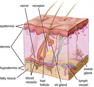
The Skin Barrier
The skin is a critical physical barrier composed of three main layers:
Epidermis: Thin, outer layer; surface cells are shed, removing microbes.
Dermis: Thick, middle layer with hair follicles, glands, nerves, and vessels.
Hypodermis: Deep, fatty layer providing cushioning and insulation.
Together, these layers prevent microbial entry into deeper tissues.
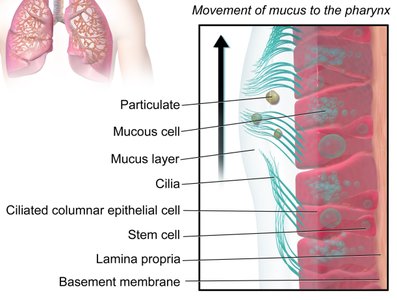
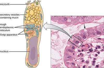
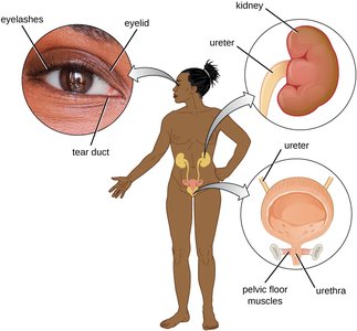
Mucous Membranes
Mucous membranes line the respiratory, digestive, urinary, and reproductive tracts. They consist of tightly joined epithelial cells that secrete mucus, trapping microbes and debris. Mechanical actions, such as the mucociliary escalator in the respiratory tract, help remove trapped pathogens.
Mucociliary escalator: Ciliated cells move mucus upward to be expelled or swallowed.
Clinical relevance: Damage to this system (e.g., by smoking or cystic fibrosis) increases infection risk.
Digestive Tract as a Physical Barrier
The digestive tract is a major entry point for microbes, but its mucous membranes and mechanical actions (e.g., peristalsis) provide strong nonspecific protection. Goblet cells secrete mucus, trapping pathogens, which are then moved and eliminated as feces.
Endothelia as Protective Barriers
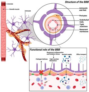
Endothelial cells line blood, lymphatic, and urogenital vessels, forming tight barriers. The blood–brain barrier is a specialized endothelium that protects the central nervous system from infection.
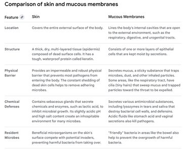
Comparison of Skin and Mucous Membranes
Feature | Skin | Mucous Membranes |
|---|---|---|
Location | Covers entire external surface | Lines internal cavities open to the environment |
Structure | Thick, multi-layered, keratinized | One or more layers, mucus-secreting |
Physical Barrier | Impermeable, shedding cells | Sticky mucus traps microbes |
Chemical Defenses | Sebum, fatty acids, low pH | Antimicrobial substances, lysozyme, acidic pH |
Resident Microbes | Beneficial skin microbiota | "Friendly" bacteria in bowel, vagina |
Mechanical Defenses
Mechanical defenses physically expel microbes from the body, preventing colonization. Examples include shedding of skin cells, mucociliary clearance, peristalsis, flushing action of urine, and tears.
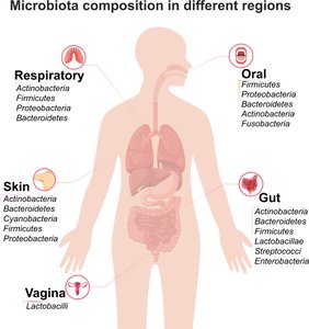
Microbiome as a Physical Defense
Resident microbiota inhabit the skin, respiratory, gastrointestinal, and urogenital tracts, competing with pathogens for binding sites and nutrients. Disruption of the microbiome (e.g., by antibiotics) can increase susceptibility to infections.
Chemical Defenses
Chemical Mediators in Innate Immunity
Chemical mediators are substances that inhibit microbial colonization and infection. They may be endogenous (produced by human cells) or exogenous (produced by microbiota). Their production can be constitutive or induced by infection.
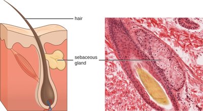
Chemical and Enzymatic Mediators on the Skin
Sebum: Secreted by sebaceous glands, coats skin and hair, sealing pores and preventing bacterial invasion.
Microbiome-derived mediators: Microbes degrade sebum, producing oleic acid, which lowers skin pH and inhibits pathogens.
Chemical Mediators Across Body Systems
Skin: Sebum, oleic acid
Digestive tract: Saliva (lactoperoxidase), stomach acid, intestinal enzymes, bile
Urinary tract: Slightly acidic urine
Female reproductive system: Lactobacilli produce lactic acid, lowering vaginal pH
Eyes: Tears contain lysozyme and lactoferrin
Ears: Cerumen (earwax) lowers pH
Respiratory tract: Lysozyme, lactoferrin, surfactant
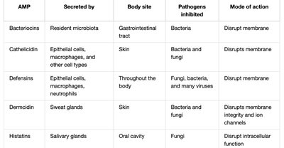
Antimicrobial Peptides (AMPs)
AMPs are small, cell-derived peptides with broad-spectrum antimicrobial activity. They can be constitutively produced or induced by pathogens. AMPs damage microbial membranes, destroy nucleic acids, or interfere with cell wall synthesis.
Defensins: Produced by epithelial cells, macrophages, neutrophils; damage microbial membranes.
Bacteriocins: Produced by resident microbiota in the gut.
AMP | Secreted by | Body site | Pathogens inhibited | Mode of action |
|---|---|---|---|---|
Bacteriocins | Resident microbiota | Gastrointestinal tract | Bacteria | Disrupt membrane |
Cathelicidin | Epithelial cells, macrophages | Skin | Bacteria, fungi | Disrupts membrane |
Defensins | Epithelial cells, macrophages, neutrophils | Throughout body | Fungi, bacteria, viruses | Disrupt membrane |
Dermcidin | Sweat glands | Skin | Bacteria, fungi | Disrupts membrane integrity |
Histatins | Salivary glands | Oral cavity | Fungi | Disrupts intracellular function |
Plasma Protein Mediators
Plasma contains key nonspecific immune proteins, including acute-phase proteins, complement proteins, and cytokines. These proteins act broadly against pathogens and support blood clotting and homeostasis.
Acute-phase proteins: Produced in response to infection or injury; enhance pathogen recognition and limit growth.
Complement proteins: Enhance pathogen elimination via lysis, opsonization, and inflammation.
Cytokines: Coordinate immune responses.
The Complement System
The complement system is a group of over 30 plasma proteins that circulate as inactive precursors. They are activated by three pathways:
Alternative pathway: Innate, nonspecific activation.
Classical pathway: Triggered by antibodies (links innate and adaptive immunity).
Lectin pathway: Triggered by mannose-binding lectin binding to microbial carbohydrates.
Activation leads to pathogen destruction through lysis, opsonization, and inflammation.
Cytokines
Cytokines are soluble proteins that mediate communication between immune cells. They regulate cell proliferation, differentiation, chemotaxis, and apoptosis. Cytokines can act in autocrine, paracrine, or endocrine fashions.
Interleukins (ILs): Regulate immune functions.
Chemokines: Recruit leukocytes to infection sites.
Interferons (IFNs): Antiviral cytokines; inhibit viral replication and activate immune cells.
Inflammation-Eliciting Mediators
Cytokines trigger the production of acute-phase proteins and other mediators (e.g., histamine, leukotrienes, prostaglandins, bradykinin) that promote inflammation, vasodilation, and increased vascular permeability, facilitating immune cell recruitment and pathogen elimination.
Cellular Defenses
Formed Elements of Blood
Blood contains three major formed elements:
Red blood cells (erythrocytes): Oxygen transport.
Platelets (thrombocytes): Blood clotting and tissue repair.
White blood cells (leukocytes): Immune defense (focus of innate immunity).
All blood cells originate from hematopoietic stem cells in the bone marrow (hematopoiesis).
Granulocytes
Neutrophils (PMNs): Phagocytose and kill bacteria; release antimicrobial granules and form neutrophil extracellular traps (NETs).
Eosinophils: Defend against protozoa and helminths; involved in allergic reactions.
Basophils: Release histamine and other mediators; important in allergy and inflammation.
Mast cells: Similar to basophils but reside in tissues; key in allergic and inflammatory responses.
Agranulocytes
Lymphocytes: Include natural killer (NK) cells (innate immunity), B cells, and T cells (adaptive immunity).
Monocytes: Differentiate into macrophages and dendritic cells, which are phagocytic and bridge innate and adaptive immunity.
Phagocytosis and Pathogen Recognition
Phagocytes (e.g., neutrophils, macrophages, dendritic cells) ingest and destroy pathogens. Recognition is enhanced by opsonization (coating with antibodies or complement) and by pattern recognition receptors (PRRs) that detect pathogen-associated molecular patterns (PAMPs).
Phagocytosis steps: Recognition, engulfment (phagosome formation), fusion with lysosome (phagolysosome), digestion, and exocytosis of debris.
Antigen presentation: Macrophages and dendritic cells display pathogen antigens to activate adaptive immunity.
Inflammatory Response
Inflammation is triggered by tissue damage or infection, leading to vasodilation, increased vascular permeability, and recruitment of immune cells. Acute inflammation is essential for pathogen elimination and tissue repair, while chronic inflammation can cause tissue damage and contribute to disease.
Five signs of inflammation: Redness, heat, swelling, pain, altered function.
Chronic inflammation: Persistent, low-level response; can form granulomas (e.g., tuberculosis).
Fever
Fever is a systemic inflammatory response that raises body temperature, enhancing immune activity and inhibiting pathogen growth. It is regulated by the hypothalamus in response to pyrogens (e.g., cytokines, bacterial toxins).
Benefits: Enhances leukocyte activity, inhibits pathogens, increases iron sequestration.
Risks: Excessive fever can cause organ damage or be fatal.
Summary Table: Chemical Defenses of Nonspecific Innate Immunity
Defense | Examples | Function |
|---|---|---|
Chemicals and enzymes in body fluids | Sebum, oleic acid, lysozyme, acid, digestive enzymes, lactoferrin, surfactant | Inhibit or kill bacteria, sequester iron, kill bacteria |
Antimicrobial peptides | Defensins, bacteriocins, dermcidin, cathelicidin, histatins | Kill bacteria by attacking membranes or interfering with cell functions |
Plasma protein mediators | Acute-phase proteins, complement proteins | Inhibit growth, opsonization, inflammation |
Cytokines | Interleukins, chemokines, interferons | Stimulate, modulate, and recruit immune cells |
Inflammation-eliciting mediators | Histamine, leukotrienes, prostaglandins, bradykinin | Promote inflammation, fever, edema |
