 Back
BackInnate Immunity: The Second Line of Defense
Study Guide - Smart Notes
 Tailored notes based on your materials, expanded with key definitions, examples, and context.
Innate Immunity: The Second Line of Defense
Overview of the Second Line of Defense
The second line of defense in innate immunity is activated when pathogens breach the skin or mucous membranes. This system is composed of various cells, antimicrobial chemicals, and physiological processes, many of which are present in or originate from the blood. These components work together to provide a rapid, nonspecific response to invading microorganisms.
Defense Components of Blood: Plasma
Plasma is the liquid portion of blood, primarily consisting of water, electrolytes, dissolved gases, nutrients, and proteins. When clotting factors are removed, the remaining fluid is called serum. Plasma contains important defense molecules such as iron-binding proteins, complement proteins, and antibodies. Iron is essential for microbial metabolism, and some microbes secrete proteins to capture iron from the host.
Defense Components of Blood: Formed Elements
The cellular components of blood, known as formed elements, include erythrocytes, platelets, and leukocytes. Each type plays a distinct role in immunity and homeostasis:
Erythrocytes: Transport oxygen and carbon dioxide.
Platelets: Involved in blood clotting and inflammation.
Leukocytes: Defend the body against invaders and are subdivided into granulocytes and agranulocytes.
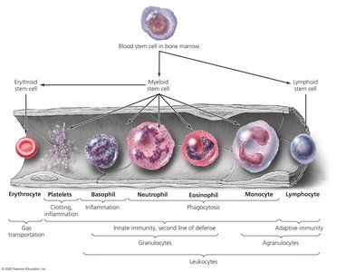
Leukocytes: Granulocytes
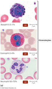
Granulocytes are leukocytes with visible granules in their cytoplasm, which stain differently with specific dyes. The three main types are:
Basophils: Stain blue with basic dyes; release inflammatory chemicals but do not phagocytize pathogens.
Eosinophils: Stain red/orange with acidic dyes; phagocytize pathogens, attack helminths, and can kill without phagocytosis.
Neutrophils: Stain lilac with a mix of dyes; phagocytize pathogens, produce antimicrobial chemicals, and form neutrophil extracellular traps (NETs).
Leukocytes: Agranulocytes
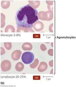
Agranulocytes have a uniform cytoplasm under light microscopy and include:
Lymphocytes: Primarily involved in adaptive immunity; natural killer (NK) lymphocytes are part of innate immunity.
Monocytes: Differentiate into dendritic cells and macrophages, which are phagocytic and ingest foreign particles.
Laboratory Analysis of Leukocytes
Laboratory evaluation of leukocyte populations, such as a differential white blood cell count, can indicate disease states:
Increased eosinophils suggest allergies or parasitic worm infection.
Bacterial infections often cause elevated leukocytes and neutrophils.
Viral infections typically increase lymphocyte numbers.
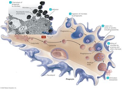
Phagocytosis: Mechanism and Stages
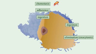
Phagocytosis is the process by which certain cells (phagocytes) ingest and destroy pathogens. The process involves six stages:
Chemotaxis: Movement of phagocytes toward chemical signals from pathogens or damaged cells.
Adhesion: Attachment of phagocyte to the pathogen.
Ingestion: Engulfment of the pathogen into a phagosome.
Maturation: Fusion of the phagosome with lysosomes to form a phagolysosome.
Killing: Destruction of the pathogen by enzymes and toxic substances.
Elimination: Expulsion of digested material by exocytosis.
Nonspecific Chemical Defenses Against Pathogens
The second line of defense also includes nonspecific chemical mechanisms:
Toll-like receptors (TLRs): Integral membrane proteins on phagocytes that recognize pathogen-associated molecular patterns (PAMPs) and trigger defensive responses such as inflammation, apoptosis, and stimulation of adaptive immunity.
NOD proteins: Cytosolic proteins that detect PAMPs and initiate inflammation and apoptosis.
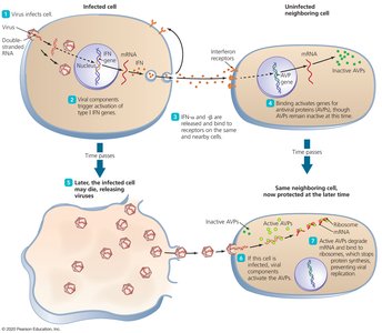
Interferons: Proteins released by host cells to slow the spread of viral infections. Type I interferons (alpha and beta) are produced early, while Type II (gamma) is produced later by T lymphocytes and NK cells.
TLR | PAMP (Microbial Molecule) |
|---|---|
TLR1 | Bacterial lipopeptides, certain parasite proteins |
TLR2 | Peptidoglycan, lipoteichoic acid, yeast cell wall |
TLR4 | Lipid A in LPS (Gram-negative bacteria) |
TLR5 | Flagellin (bacterial flagella) |
TLR6 | Bacterial lipopeptides, lipoteichoic acid, yeast cell wall |
TLR10 | Unknown component of influenzaviruses |
TLR3 | Double-stranded RNA (viruses) |
TLR7/8 | Single-stranded viral RNA |
TLR9 | Unmethylated CpG DNA (viral/bacterial) |
Summary Table: Leukocyte Types and Functions
Cell Type | Main Function | Granules Present? |
|---|---|---|
Erythrocyte | Gas transport | No |
Platelet | Clotting, inflammation | No |
Basophil | Inflammation | Yes |
Neutrophil | Phagocytosis, innate immunity | Yes |
Eosinophil | Phagocytosis, helminth defense | Yes |
Monocyte | Phagocytosis (as macrophage/dendritic cell) | No |
Lymphocyte | Adaptive immunity | No |
Key Points and Examples
Phagocytosis is essential for destroying pathogens; pathogens are killed in phagolysosomes by acid and oxidative radicals.
Gamma interferon is produced late in infection by T lymphocytes and NK cells.
Toll-like receptors do not target eukaryotic flagellar proteins, but do recognize bacterial flagellin, lipid A, and viral RNA.
