 Back
Introduction to Bacteria: Structure, Classification, and Laboratory Identification
Study Guide - Smart Notes
 Tailored notes based on your materials, expanded with key definitions, examples, and context.
Introduction to Bacteria
Scope of Medical Microbiology
Medical microbiology encompasses the study of various microorganisms that impact human health, including bacteria, fungi, parasites, and viruses. Each group is studied within its own discipline, such as bacteriology (bacteria), mycology (fungi), parasitology (eukaryotic microbes), and virology (viruses and prions).
Bacteria: Prokaryotic, unicellular organisms with diverse shapes and metabolic capabilities.
Fungi: Includes yeasts and molds, eukaryotic organisms.
Parasites: Protozoa, helminths, and ectoparasites, all eukaryotic.
Viruses: Acellular infectious agents, sometimes including prions.
Prokaryotic vs. Eukaryotic Cells
Key Differences
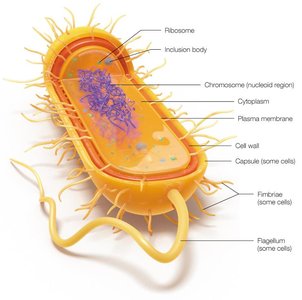
Bacteria are prokaryotic organisms, meaning they lack a nucleus and membrane-bound organelles. They are typically microscopic, unicellular, and possess a cell wall (except for Mycoplasma species).
Prokaryotes: No nucleus, no organelles, single circular chromosome, cell wall present.
Eukaryotes: Nucleus present, organelles present, multiple linear chromosomes, cell wall may be present (in fungi and plants).
Bacterial Taxonomy and Classification
Species and Strains
Bacterial taxonomy classifies organisms based on genetic, biochemical, and phenotypic criteria. A species in bacteriology is defined by genetic similarity and observable characteristics, while a strain refers to genetic variants within a species.
Species: Group of organisms with similar genetic and phenotypic traits.
Strain: Subgroup within a species, often with unique features (e.g., pathogenicity, metabolism).
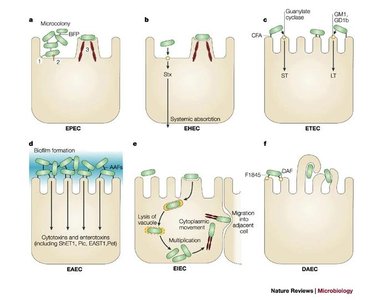
Example: Escherichia coli (species) includes multiple strains such as EPEC, EHEC, ETEC, EAEC, EIEC, and DAEC, each with distinct pathogenic mechanisms.
Bacterial Morphology
Colony and Cellular Morphology
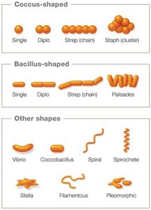
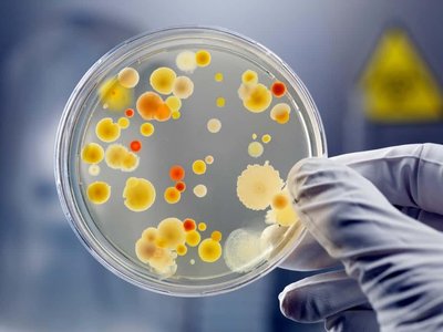
Bacterial morphology refers to the form and structure of bacteria, both at the colony and cellular level. Colony morphology includes macroscopic traits such as size, shape, color, elevation, texture, and margin. Cellular morphology includes microscopic traits such as cell shape and arrangement.
Colony Morphology: Observed on solid media; useful for preliminary identification.
Cellular Morphology: Observed under a microscope; includes shape (bacilli, cocci, vibrio, spirochetes, etc.) and arrangement (chains, clusters, pairs).
Bacterial Growth and Division
Binary Fission
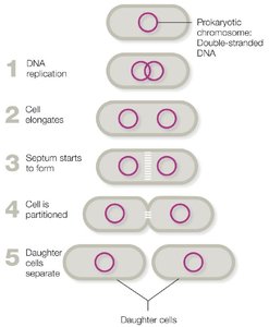
Bacteria reproduce asexually by binary fission, a process in which a single cell divides into two genetically identical daughter cells. This leads to rapid population growth under favorable conditions.
Steps of Binary Fission:
DNA replication
Cell elongation
Septum formation
Partitioning of the cell
Separation of daughter cells
Consequence: Short generation time and genetic uniformity among progeny.
Laboratory Identification of Bacteria
Methods of Detection
Several laboratory techniques are used to identify bacteria in clinical samples:
Microscopy: Observes cell shape, size, arrangement, and staining properties.
Culturing: Grows bacteria on artificial media to observe colony morphology and metabolic traits.
Immunologic Methods: Detects bacterial antigens or host antibodies.
Molecular Methods: Detects bacterial DNA using techniques like PCR.
Bacterial Cell Wall Structure
Gram-Positive vs. Gram-Negative Cell Walls
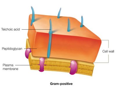
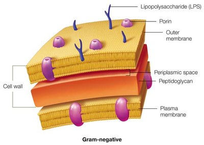
The bacterial cell wall provides structural support and protection. Its composition is the basis for the Gram stain, which differentiates bacteria into Gram-positive and Gram-negative groups.
Gram-Positive: Thick peptidoglycan layer, no outer membrane, stains purple.
Gram-Negative: Thin peptidoglycan layer, outer membrane with lipopolysaccharide (LPS), stains pink/red.
Gram Stain Procedure
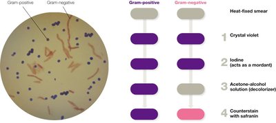
The Gram stain is a differential staining technique that classifies bacteria based on cell wall structure.
Crystal violet (primary stain)
Iodine (mordant)
Alcohol/acetone (decolorizer)
Safranin (counterstain)
Gram-positive cells retain crystal violet and appear purple; Gram-negative cells lose crystal violet and take up safranin, appearing pink.
Specialized Bacterial Structures
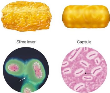
Capsule
The capsule is an extracellular layer found in some bacteria. It is composed mainly of carbohydrates and serves as a protective barrier, aiding in adherence and resistance to desiccation and immune defenses.
Function: Adherence, protection from drying, evasion of immune response.
Composition: Polysaccharides (carbohydrates).
Flagella
Flagella are protein-based structures that provide motility to bacteria. They are not present in all species and can be arranged in various patterns (monotrichous, amphitrichous, lophotrichous, peritrichous).
Function: Motility, movement toward favorable environments or within host tissues.
Composition: Protein (flagellin).

Fimbriae and Pili
Fimbriae and pili are proteinaceous appendages involved in adherence and, in the case of pili, genetic exchange (conjugation).
Fimbriae: Short, bristle-like fibers for adherence to surfaces.
Pili: Longer, more rigid structures; some (sex pili) facilitate DNA transfer between cells.
Endospores
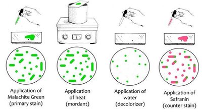
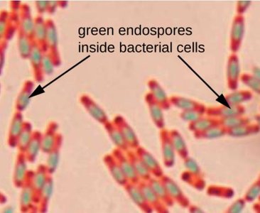
Endospores are highly resistant, dormant structures formed by certain genera (e.g., Bacillus, Clostridium) under adverse conditions. They are extremely difficult to destroy and require autoclaving or sporicidal agents for inactivation.
Function: Survival under harsh conditions (heat, desiccation, chemicals).
Clinical significance: Endospore-forming bacteria are major concerns in healthcare due to their resistance.
Bacterial Adaptations and Pathogenicity
Adaptations for Survival and Virulence
Bacterial structures such as flagella, capsules, pili, and fimbriae are adaptations that enhance survival in diverse environments. In the context of a human host, these adaptations can contribute to pathogenicity (the ability to cause disease).
Flagella: Movement toward or within host tissues.
Capsule: Evasion of phagocytosis and immune defenses.
Fimbriae/Pili: Attachment to host cells and tissues.
Biofilm-associated structures: Persistent colonization and increased resistance to host defenses.
Virulence is the degree to which a pathogen can cause disease. Not all bacteria with these structures are pathogens.
Summary Table: Medically Relevant Bacterial Genera
Gram Positive | Gram Negative | Other |
|---|---|---|
Staphylococcus (cocci) Streptococcus (cocci) Enterococcus (cocci) Bacillus* (bacilli) Corynebacteria (bacilli) Clostridium/Clostridioides* (bacilli) Cutibacterium* (bacilli) Listeria (bacilli) | Neisseria (cocci) Bacteroides* (bacilli) Bordetella (bacilli) Campylobacter (bacilli) Enterobacteriaceae (bacilli) Helicobacter (bacilli) Haemophilus (bacilli) Legionella (bacilli) Pseudomonas (bacilli) Vibrio (bacilli) | Mycobacteriumα Nocardiaα BorreliaƧ Chlamydia Mycoplasma Rickettsia TreponemaƧ |
* indicates anaerobe; α indicates acid-fast; • indicates endospore formation; Ƨ indicates spirochete
Key Learning Objectives
Differentiate between prokaryotic and eukaryotic microbes.
Define: strain, morphology, endospore.
Describe the various shapes and arrangements of bacteria.
Compare and contrast Gram-positive and Gram-negative bacteria.
Discuss the function and composition of bacterial cell walls, capsules, flagella, fimbriae, pili, and endospores.
Explain the significance of endospore-forming bacteria in healthcare settings.
Describe the Gram stain procedure and expected results.
Describe the common laboratory techniques used to identify microbes in patient samples.
