 Back
BackIntroduction to Microbiology and Functional Anatomy of Prokaryotic Cells
Study Guide - Smart Notes
 Tailored notes based on your materials, expanded with key definitions, examples, and context.
Introduction to Microbiology
What are Microorganisms?
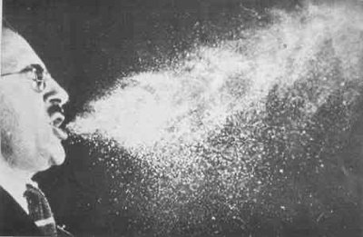
Microorganisms, or microbes, are organisms that are too small to be seen with the unaided eye. They are found in almost every environment on Earth and play essential roles in ecosystems and human health.
Definition: Microorganisms include bacteria, archaea, fungi, protozoa, algae, and viruses.
Visibility: Cannot be seen with the naked eye; require a microscope for observation.
Where are Microorganisms Found?
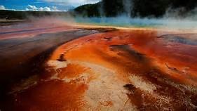
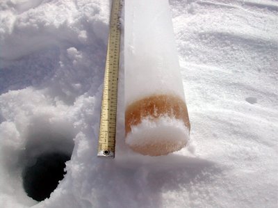
Microbes inhabit a vast range of environments, from common to extreme, and are even found in and on the human body.
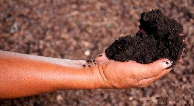
Common Environments: Water, soil, air, and on living hosts.
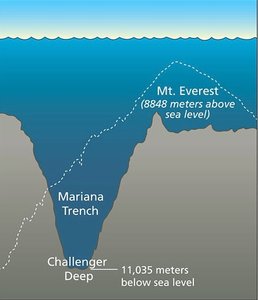
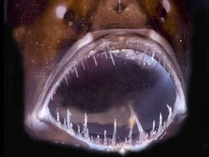
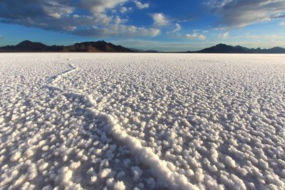
Extreme Environments: Polar ice caps, hot springs, ocean depths, volcanic soils, and salt flats.
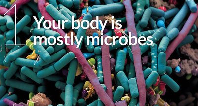
Human Body: Microbes outnumber human cells and are essential for digestion and immunity.


Classification of Microorganisms
The classification of microorganisms is based on cellular organization and genetic sequencing. Carl Woese developed the three-domain system in 1978.
Three Domains:
Bacteria: Prokaryotic, peptidoglycan cell walls.
Archaea: Prokaryotic, lack peptidoglycan, often extremophiles.
Eukarya: Eukaryotic, includes protists, fungi, plants, and animals.
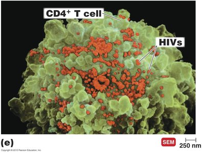
Viruses: Acellular, not classified as living cells, require host cells for replication.
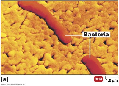
Types of Microorganisms
Bacteria: Prokaryotes, peptidoglycan cell walls, reproduce by binary fission, diverse metabolism.
Archaea: Prokaryotes, lack peptidoglycan, live in extreme environments, non-pathogenic.
Fungi: Eukaryotes, chitin cell walls, absorb organic chemicals, include yeasts (unicellular) and molds/mushrooms (multicellular).

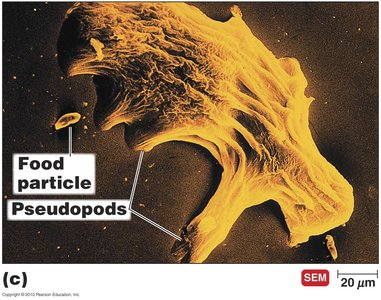
Protozoa: Eukaryotes, absorb/ingest organic chemicals, motile via pseudopods, cilia, or flagella.
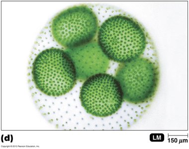
Algae: Eukaryotes, cellulose cell walls, photosynthetic, produce oxygen, non-pathogenic.
Viruses: Acellular, DNA or RNA core, protein coat, replicate only in host cells.
Historical Discoveries in Microbiology
Early observations and experiments laid the foundation for microbiology as a science.
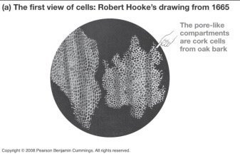
Robert Hooke (1665): Observed cells in cork, leading to cell theory.
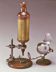
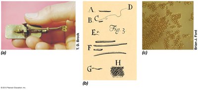
Anton van Leeuwenhoek (1673-1723): First to describe live microorganisms, called them "animalcules."
Spontaneous Generation vs. Biogenesis
The debate over the origin of life led to key experiments disproving spontaneous generation.
Spontaneous Generation: Hypothesis that life arises from nonliving matter.
Biogenesis: Hypothesis that living cells arise only from preexisting living cells.
Key Experiments:
Francesco Redi (1668): Showed maggots come from flies, not spontaneous generation.
John Needham (1745): Claimed microbes developed spontaneously in broth.
Lazzaro Spallanzani (1765): Showed no growth in sealed, boiled broth.
Louis Pasteur (1861): Demonstrated microbes are present in the air and disproved spontaneous generation with swan-neck flask experiments.
Importance of Microorganisms
Microorganisms have profound impacts on human health, agriculture, industry, and the environment.
Human Health: Cause infectious diseases but also aid in digestion and immunity.
Agriculture: Involved in nutrient cycles (nitrogen, sulfur), plant growth, and animal nutrition.
Industry: Used in food production, antibiotics, biotechnology, bioremediation, and biofuels.
Functional Anatomy of Prokaryotic Cells
Prokaryotes vs. Eukaryotes
Prokaryotic and eukaryotic cells differ in structure and function.
Prokaryotes: No nucleus, single circular chromosome, 70S ribosomes, no organelles, peptidoglycan (bacteria) or pseudomurein (archaea) cell walls, binary fission.
Eukaryotes: Nucleus, paired chromosomes, 80S ribosomes, organelles, polysaccharide cell walls (plants/fungi), mitosis.
Bacterial Cell Size and Shape
Bacteria exhibit a variety of sizes and shapes, which are important for identification and classification.
Size: Typically 0.2–1.0 µm wide and 2–8 µm long.
Shapes:
Bacillus: Rod-shaped
Coccus: Spherical
Spiral: Includes vibrio (curved rod), spirillum (rigid spiral), spirochete (flexible spiral)
Other: Star-shaped, rectangular, pleomorphic (variable shape)
Bacterial Cell Structure
Bacterial cells have several essential structures, each with specific functions.
Plasma Membrane: Phospholipid bilayer with proteins, selectively permeable, site of ATP production, contains hopanoids (bacteria) or sterols (eukaryotes/mycoplasmas).
Cell Wall: Provides shape and protection, composed of peptidoglycan in bacteria.
Cytoplasm: Contains ribosomes, DNA, and inclusions.
External Structures: Capsule, flagella, pili, fimbriae (involved in motility, attachment, and virulence).
Plasma Membrane: Structure and Function
The plasma membrane is a dynamic structure essential for cell survival.
Fluid Mosaic Model: Lipid bilayer with embedded proteins; flexible and self-sealing.
Functions:
Encloses cytoplasm
Selective permeability
ATP production (electron transport chain)
Photosynthetic pigments (chromatophores)
Anchors external structures
Movement Across the Plasma Membrane
Substances move across the membrane by passive or active processes.
Passive Processes: No energy required; includes simple diffusion, facilitated diffusion, and osmosis.
Active Processes: Require energy (ATP); includes active transport and group translocation.
Osmosis and Tonicity
Osmosis is the movement of water across a selectively permeable membrane. Tonicity describes the effect of solute concentration on cell volume.
Isotonic: Equal solute concentration inside and outside; no net water movement.
Hypotonic: Lower solute outside; water enters cell, may cause lysis.
Hypertonic: Higher solute outside; water leaves cell, causing shrinkage (plasmolysis).
Bacterial Cell Wall: Structure and Function
The cell wall provides structural support and protection against osmotic lysis.
Peptidoglycan: Polymer of N-acetylglucosamine (NAG) and N-acetylmuramic acid (NAM) linked by β 1-4 glycosidic bonds and cross-linked by peptides.
Gram-Positive: Thick peptidoglycan, teichoic acids, no outer membrane.
Gram-Negative: Thin peptidoglycan, outer membrane with lipopolysaccharide (LPS), periplasmic space.
Mycobacteria: Waxy cell wall with mycolic acids, slow growth, resistant to desiccation and digestion.
Atypical: Mycoplasmas lack cell wall; Archaea have pseudomurein or S-layer.
Gram Staining
Gram staining differentiates bacteria based on cell wall structure.
Gram-Positive: Retain crystal violet stain (purple) due to thick peptidoglycan.
Gram-Negative: Lose crystal violet, take up safranin (pink/red) due to thin peptidoglycan and outer membrane.
Damage to the Cell Wall
Certain antibiotics (e.g., penicillin, vancomycin) and enzymes (e.g., lysozyme) target the cell wall, leading to cell lysis.
Penicillin: Inhibits cross-bridge formation in peptidoglycan (mainly Gram-positive).
Lysozyme: Breaks glycosidic bonds in peptidoglycan.
Summary Table: Comparison of Cell Wall Types
Feature | Gram-Positive | Gram-Negative | Mycobacteria | Mycoplasma | Archaea |
|---|---|---|---|---|---|
Peptidoglycan | Thick | Thin | Present, with mycolic acids | Absent | Pseudomurein or S-layer |
Outer Membrane | Absent | Present (LPS) | Absent | Absent | Absent |
Teichoic Acids | Present | Absent | Absent | Absent | Absent |
Mycolic Acids | Absent | Absent | Present | Absent | Absent |
Sensitivity to Penicillin | High | Low | Variable | None | None |
Additional info: This guide covers foundational concepts from Chapters 1 and 4 of a typical college microbiology course, including the diversity, classification, and structure of microorganisms, as well as the functional anatomy of prokaryotic cells.
