 Back
BackIntroduction to Microbiology and Functional Anatomy of Prokaryotic Cells
Study Guide - Smart Notes
 Tailored notes based on your materials, expanded with key definitions, examples, and context.
Introduction to Microbiology
What are Microorganisms?
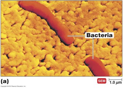
Microorganisms, or microbes, are organisms that are too small to be seen with the unaided eye. They are found in nearly every environment on Earth and play essential roles in ecosystems, human health, and industry.
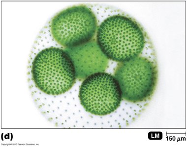
Definition: Microorganisms include bacteria, archaea, fungi, protozoa, algae, and viruses.
Size: Typically measured in micrometers (µm) or nanometers (nm).
Where Do You Find Microorganisms?
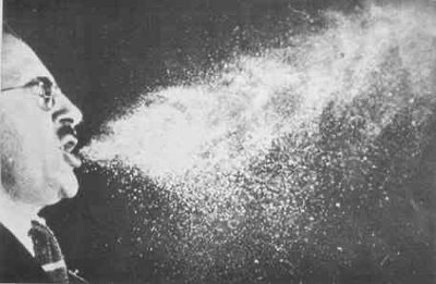
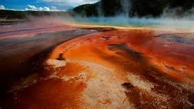
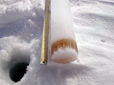
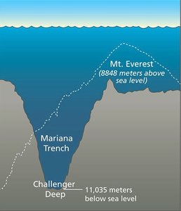
Microbes inhabit a vast range of environments, from common to extreme.
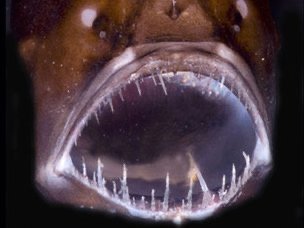
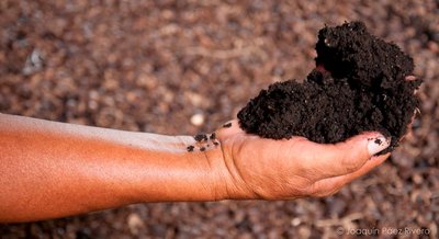
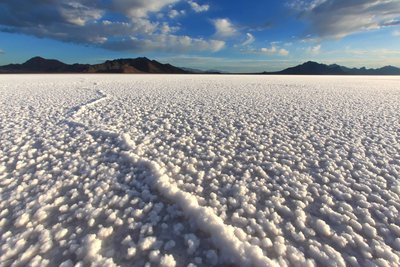
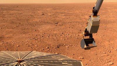
Common environments: Water, soil, air, and even the human body.
Extreme environments: Polar ice caps, hot springs, ocean depths, volcanic soil, and salt flats.


Classification of Microorganisms
The classification of organisms was revolutionized by Carl Woese in 1978, who proposed three domains based on cellular organization:
Bacteria: Prokaryotic, peptidoglycan cell walls, diverse metabolism.
Archaea: Prokaryotic, lack peptidoglycan, often extremophiles.
Eukarya: Eukaryotic, includes protists, fungi, plants, and animals.
Types of Microorganisms
Prokaryotic: Bacteria, Archaea
Eukaryotic: Fungi, Protozoa, Algae
Acellular: Viruses
Bacteria
Prokaryotes with peptidoglycan cell walls
Reproduce by binary fission
Metabolically diverse: use organic/inorganic chemicals or photosynthesis
Includes pathogens
Archaea
Prokaryotes lacking peptidoglycan
Live in extreme environments (halophiles, thermophiles)
None are known pathogens
Fungi
Eukaryotes with chitin cell walls
Use organic chemicals for energy
Molds and mushrooms are multicellular; yeasts are unicellular
Some are pathogens
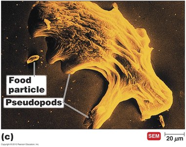
Protozoa
Eukaryotes, absorb or ingest organic chemicals
Motile via pseudopods, cilia, or flagella
Many are human pathogens
Algae
Eukaryotes, cellulose cell walls
Photosynthetic, produce oxygen and organic compounds
None are pathogenic
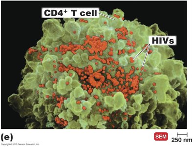
Viruses
Acellular, consist of DNA or RNA core surrounded by a protein coat
May have a lipid envelope
Replicate only inside living host cells
Early Discoveries in Microbiology
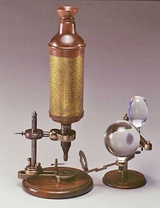
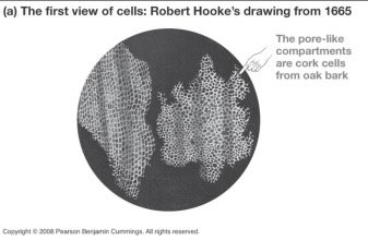
1665: Robert Hooke observed cells in cork tissue.
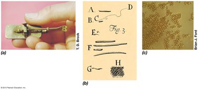
1673-1723: Anton van Leeuwenhoek described live microorganisms, calling them "animalcules."
The Debate Over Spontaneous Generation
Historically, scientists debated whether life could arise spontaneously from nonliving matter (spontaneous generation) or only from preexisting life (biogenesis).
Francesco Redi (1668): Showed that maggots on meat came from fly eggs, not spontaneous generation.
John Needham (1745): Claimed microbes developed spontaneously in boiled broth.
Lazzaro Spallanzani (1765): Showed that sealed, boiled broth did not develop microbes, supporting biogenesis.
Louis Pasteur (1861): Demonstrated that microorganisms are present in the air and do not arise spontaneously.
Theory of Biogenesis
Pasteur's experiments with swan-neck flasks provided strong evidence for biogenesis, showing that sterilized broth remained free of microbes unless exposed to air containing dust and microorganisms.
The Golden Age of Microbiology
Pasteurization: Pasteur showed that spoilage bacteria could be killed by heat without damaging beverages, leading to the process of pasteurization.
Importance of Microorganisms
Microorganisms can be both beneficial and harmful.
Most are beneficial, playing roles in nutrient cycling, food production, and biotechnology.
Pathogens are microbes that cause disease.
Functional Anatomy of Prokaryotic Cells
Prokaryotes vs. Eukaryotes
Prokaryotes: One circular chromosome, no membrane-bound nucleus, no organelles, peptidoglycan cell walls (bacteria), 70S ribosomes, binary fission.
Eukaryotes: Paired chromosomes in a nuclear membrane, organelles, polysaccharide cell walls (plants/fungi), 80S ribosomes, mitosis.
Bacterial Cell Size and Shape
Size: 0.2–1.0 µm × 2–8 µm
Shapes: Bacillus (rod), Coccus (spherical), Spiral (vibrio, spirillum, spirochete), star-shaped, rectangular
Most bacteria are monomorphic; some are pleomorphic.
Bacterial Cell Wall
Peptidoglycan: Polymer of N-acetylglucosamine (NAG) and N-acetylmuramic acid (NAM) linked by β 1-4 glycosidic bonds and cross-linked by peptides.
Gram-positive: Thick peptidoglycan, teichoic acids, no outer membrane.
Gram-negative: Thin peptidoglycan, outer membrane with lipopolysaccharides, periplasmic space.
Mycobacteria: Waxy cell wall with mycolic acids, slow-growing, resistant to desiccation and digestion.
Mycoplasmas: Lack cell wall, have sterols in plasma membrane.
Archaea: May lack cell wall or have walls of pseudomurein or S-layer proteins.
Plasma Membrane
Structure: Phospholipid bilayer with proteins (fluid mosaic model), contains hopanoids (bacteria) or sterols (eukaryotes/mycoplasmas).
Functions: Selective permeability, ATP production (electron transport chain), anchoring flagella/pili/fimbriae, sometimes photosynthetic pigments.
Transport Across Membranes
Passive processes: Simple diffusion, facilitated diffusion, osmosis.
Active processes: Active transport (requires ATP), group translocation (substance chemically modified during transport).
External Structures
Glycocalyx: Capsule (organized, protective), slime layer (unorganized, loose), aids in attachment and protection.
Flagella: Motility, chemotaxis, composed of flagellin, powered by proton motive force.
Fimbriae: Attachment to surfaces.
Pili: Attachment, twitching/gliding motility, DNA transfer (sex pili).
Internal Structures
Nucleoid: Circular DNA, supercoiled, sometimes with plasmids (extra-chromosomal DNA).
Ribosomes: 70S in prokaryotes, site of protein synthesis.
Cytoskeleton: Maintains cell shape, aids in division.
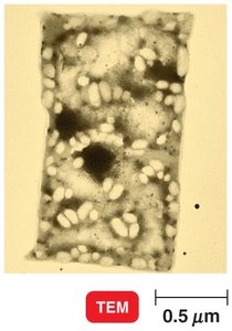
Inclusions: Storage granules (e.g., polyhydroxyalkanoate, phosphate, sulfur), carboxysomes, gas vesicles, magnetosomes.
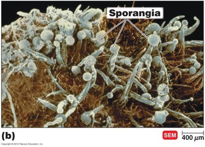
Endospores: Dormant, highly resistant structures formed by Bacillus and Clostridium; survive extreme conditions, germinate when favorable.
Summary Table: Differences Between Gram-Positive, Gram-Negative, and Mycobacterial Cell Walls
Feature | Gram-Positive | Gram-Negative | Mycobacteria |
|---|---|---|---|
Peptidoglycan | Thick | Thin | Thin, with mycolic acids |
Teichoic acids | Present | Absent | Absent |
Outer membrane | Absent | Present | Absent |
Mycolic acids | Absent | Absent | Present |
Lipid content | Low | High (LPS) | Very high |
Additional info: These notes provide a foundational overview of microbiology and the structure of prokaryotic cells, suitable for exam preparation and further study in microbial physiology, genetics, and applied microbiology.
