 Back
BackIntroduction to Microbiology & Microscopy: Foundations and Diversity Unit 1
Study Guide - Smart Notes
 Tailored notes based on your materials, expanded with key definitions, examples, and context.
Introduction to Microbiology
Definition and Scope of Microbiology
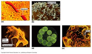
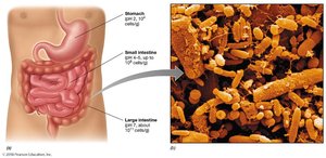
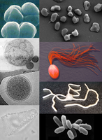
Microbiology is the study of microorganisms (microbes), which are life forms too small to be seen by the naked eye. These organisms are highly diverse in form and function, inhabiting every environment that supports life and often living in complex communities. Microbes constitute a major fraction of Earth's biomass, with an estimated 2 x 1030 cells present globally.
Microbe: A microscopic organism, including bacteria, archaea, fungi, protozoa, algae, and viruses.
Culture: Cells grown in or on a nutrient medium.
Colony: A visible mass of microbial growth, all derived from a single mother cell.
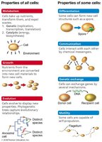
Common Structures and Activities of Cells
All cells share fundamental features and activities that define life. These include the presence of chromosomes composed of DNA, a cell membrane, cytosol, and ribosomes. Cells carry out essential functions such as growth, reproduction, evolution, metabolism, and are composed of cells.
Metabolism: Uptake of nutrients, transformation, and expulsion of wastes.
Growth: Nutrients from the environment are converted into new cell materials.
Evolution: Cells evolve to display new properties.
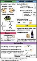
Impact of Microorganisms on Human Society
Microbial Roles and Applications
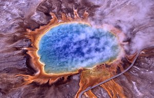
Microorganisms are the oldest forms of life, appearing approximately 3.8–4.3 billion years ago. They are known as extremophiles for their ability to inhabit extreme environments. Microbes play crucial roles in oxygen production, disease causation, biotechnology, agriculture, food production, biofuels, pharmaceuticals, and environmental cleanup.
Pathogens: Microbes that cause disease, but also serve as models for research and biotechnology.
Biofuels: Microbes are used in the production of renewable energy sources.
Bioremediation: Microbes help clean up pollutants.
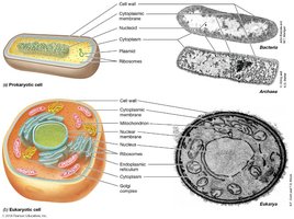
Cell Structure: Prokaryotes vs. Eukaryotes
Comparing Cell Types
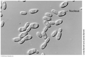
Cells are classified as either prokaryotic or eukaryotic. Prokaryotes lack a nucleus and membrane-bound organelles, are generally smaller, and include bacteria and archaea. Eukaryotes possess a nucleus and other organelles, are typically larger, and include plants, animals, fungi, and protists.
Prokaryote: "Pre-nucleus"; no nucleus or membrane-bound organelles.
Eukaryote: "True nucleus"; has nucleus and organelles.
Microscopy: Tools for Studying Microbes
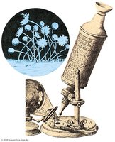
History and Principles of Microscopy
The field of microbiology began with the invention of the microscope. Robert Hooke and Antoni van Leeuwenhoek were pioneers, with Leeuwenhoek being the first to observe bacteria. Microscopy relies on magnification and resolution to visualize microorganisms.
Magnification: Ability to enlarge the image of an object.
Resolution: Ability to distinguish two adjacent objects as separate.
Limit of resolution for light microscopes: 0.2 μm.
Types of Microscopes
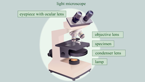
Different microscopes offer varying magnification and resolution capabilities. Light microscopes are suitable for viewing cell shapes and live cells, while electron microscopes (TEM and SEM) provide greater magnification and resolution for detailed cellular and molecular structures.
Light Microscope: Up to 1000X magnification, 0.2 μm resolution.
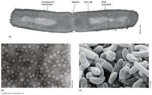
TEM (Transmission Electron Microscope): Up to 1,000,000X, 0.2 nm resolution.
SEM (Scanning Electron Microscope): Up to 100,000X, 3D imaging of surfaces.
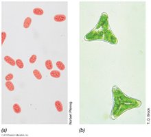

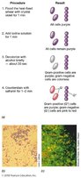
Staining Techniques in Light Microscopy
Contrast is essential for distinguishing microorganisms from their surroundings. Staining methods include simple staining (single stain) and differential staining (multiple stains, highlighting structural differences).
Simple Staining: All cells appear the same color.
Differential Staining: Cells take on different colors based on structural differences.

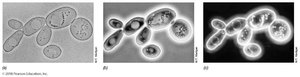
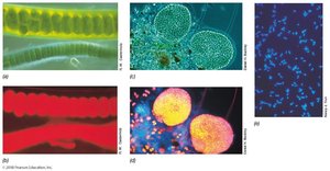
Advanced Light Microscopy Techniques
Several specialized light microscopy methods enhance visualization of live, unstained cells or specific cellular features:
Phase-contrast Microscopy: Amplifies differences in refractive index for improved contrast.
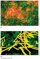
Dark-field Microscopy: Only scattered light reaches the lens, excellent for viewing motility.
Fluorescence Microscopy: Visualizes specimens that emit light after illumination; used in diagnostics and ecology.
DIC Microscopy: Uses polarized light for 3D-like appearance of internal structures.
Confocal Scanning Laser Microscopy: Generates 3D images using laser and computer, useful for thick specimens.

Historical Figures in Microbiology
Pioneers and Their Contributions
Several scientists have profoundly impacted microbiology:
Robert Hooke: First to describe cells.
Antoni van Leeuwenhoek: First to observe bacteria.
Louis Pasteur: Disproved spontaneous generation, developed pasteurization.
Ignaz Semmelweis: Advocated handwashing to prevent disease.
Robert Koch: Developed germ theory of disease, Koch's postulates.
Joseph Lister: Introduced antiseptic techniques.
Martinus Beijerinck: Discovered viruses and enrichment culture techniques.
Sergei Winogradsky: Studied microbial ecology and chemolithotrophy.
Classification: The Three Domains of Life
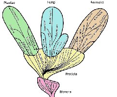
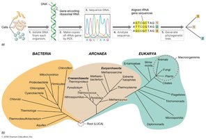
Whittaker and Woese Trees of Life
Early classification grouped all prokaryotes as Monera. Advances in molecular biology, especially rRNA sequencing, led Carl Woese to propose the Three Domains of Life: Bacteria, Archaea, and Eukarya. rRNA is universal, highly conserved, and functionally constant, making it ideal for phylogenetic studies.
Bacteria
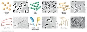
Bacteria are prokaryotes with peptidoglycan cell walls. They are unicellular but vary widely in form, with 30 major phylogenetic lineages and diverse physiologies.
Archaea
Archaea are prokaryotes with unique cell wall structures (S-layers or pseudopeptidoglycan). They are highly diverse, often associated with extreme environments, and lack known pathogens of plants, animals, or humans.

Eukarya
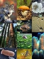
Eukarya includes all eukaryotic organisms, some unicellular and some multicellular. Eukaryotes may have cell walls (but never peptidoglycan), and are classified into at least six kingdoms, varying dramatically in size, form, and physiology.
Viruses: Unique Biological Entities
Characteristics of Viruses
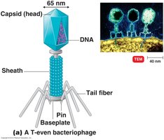
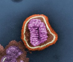
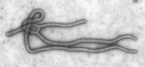
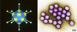
Viruses are obligate intracellular parasites and are considered non-living. They are not made of cells, cannot reproduce outside a host, and are structurally simple. Viral genomes may be DNA or RNA, protected by a protein capsid, and some have a lipid envelope. Viruses infect all three domains of life and are as diverse as their hosts.
Summary Table: Comparison of Cell Types
Feature | Prokaryote | Eukaryote |
|---|---|---|
Nucleus | No | Yes |
Membrane-bound organelles | No | Yes |
Cell wall composition | Peptidoglycan (Bacteria), S-layer/pseudopeptidoglycan (Archaea) | Varies, never peptidoglycan |
Size | Smaller | Larger |
Domains | Bacteria, Archaea | Eukarya |
Conclusion
Microbiology is a foundational discipline for understanding life at the microscopic level. The diversity, structure, and function of microbes, along with the tools used to study them, underpin many advances in science, medicine, and industry. The classification of life into three domains, the historical development of the field, and the unique nature of viruses are central concepts for any student of microbiology.
