 Back
Introduction to Microbiology: Cell Types, Pathogens, and Microbial Techniques
Study Guide - Smart Notes
 Tailored notes based on your materials, expanded with key definitions, examples, and context.
Introduction to Microbiology
Prokaryotic vs. Eukaryotic Cells
Microbiology distinguishes between prokaryotic and eukaryotic cells, which differ in structure and complexity.
Prokaryotic cells: Lack a nucleus and organelles; size ranges from 1–10 µm.
Eukaryotic cells: Possess a nucleus and organelles; size ranges from 10–100 µm.
Bacteria and Archaea are prokaryotes, while Fungi, Protists, and Helminths are eukaryotes.
Bacteria: Unicellular, contain peptidoglycan in cell walls.
Archaea: Unicellular, lack peptidoglycan, often inhabit extreme environments.
Fungi: Unicellular (yeasts) or multicellular (molds), cell walls contain chitin.
Protists: Unicellular (e.g., amoeba) or multicellular (e.g., algae).
Helminths: Multicellular parasitic worms.
Pathogens and Disease
Pathogens and Opportunistic Pathogens
Pathogens are microbes that cause disease. Only a small fraction of microbes are pathogenic.
~1,400 known pathogens; less than 1% of all microbes are pathogenic.
Opportunistic pathogens cause disease only in weakened hosts.
Historical Figures in Microbiology
Robert Hooke: First to publish descriptions of cells (1665).
Antonie van Leeuwenhoek: Refined microscopes, first to observe bacteria.
Francesco Redi: Demonstrated biogenesis with meat and maggots experiments.
Louis Pasteur: Disproved spontaneous generation, developed pasteurization, and vaccines for anthrax and rabies.
Germ Theory of Disease
The germ theory states that microbes cause infectious diseases. Key contributors include Pasteur and Koch.
Pasteur: Linked microbes to spoilage and disease; developed pasteurization.
Koch: Developed staining techniques, media for bacterial isolation, and Koch's postulates.
Koch’s Postulates of Disease
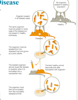
Koch's postulates are a set of criteria used to establish a causative relationship between a microbe and a disease:
The same microbe must be present in every case of the disease.
The organism must be isolated from the diseased host and grown as a pure culture.
The isolated organism should cause the same disease when inoculated into a susceptible host.
The organism must then be re-isolated from the inoculated, diseased animal.
Aseptic Techniques and Hand Hygiene
Development of Aseptic Techniques
Aseptic techniques are essential for preventing healthcare-acquired infections (HAIs) and limiting disease spread.
Ignaz Semmelweis: Recommended hand washing to reduce childbed fever mortality (1840s).
Joseph Lister: Used phenol disinfectant, sterilized instruments, and sanitized wounds to prevent infection.
Aseptic processes include sterile media, instruments, decontaminated surfaces, and protective clothing.
Microbial Classification and Taxonomy
Taxonomic Hierarchy
Microbes are classified using a hierarchical system established by Carl Linnaeus:
Binomial nomenclature: Genus (capitalized) and species (lowercase), italicized (e.g., Escherichia coli).
Taxonomic ranks: Domain, Kingdom, Phylum, Class, Order, Family, Genus, Species.
Domains are the broadest grouping: Bacteria, Archaea, and Eukarya.
Bacteria: Unicellular, prokaryotic.
Archaea: Unicellular, prokaryotic, often extremophiles, no known pathogens.
Eukarya: Unicellular and multicellular eukaryotes (Animalia, Plant, Fungal, Protist).
Species and Strains
Eukaryotic species: Organisms that can sexually reproduce together.
Prokaryotic species: Cells sharing physical characteristics and at least 70% DNA similarity; 97% identical 16S rRNA sequence.
Strain: Genetic variants of the same species, often denoted by numbers/letters (e.g., E. coli K-12).
Microbial Relationships and Biofilms
Symbiotic Relationships
Microbes interact with hosts in various symbiotic relationships:
Parasitism: Microbe harms the host (e.g., pathogens).
Mutualism: Both microbe and host benefit.
Commensalism: Microbe benefits; host is unaffected.
Biofilms
Biofilms are sticky communities of microbes that form protective matrices on surfaces.
Biofilms can develop on teeth, contact lenses, water filters, cutting boards, and catheters.
NIH estimates 60–80% of human infectious diseases are due to biofilm-forming microbes.
Biofilms are resistant to antibiotics and immune responses.
Biofilm formation steps:
Cells seed a biofilm and produce adhesion factors.
Secrete sticky substances forming a protective matrix.
Bacteria grow in layers; microbes are periodically released as planktonic cells.
Normal Microbiota and Its Functions
Normal Microbiota
Normal microbiota (flora) includes bacteria, archaea, and eukaryotic microbes that inhabit the human body.
Functions: Train immune system, produce vitamins, aid digestion, influence mood and brain function.
Development influenced by delivery method and feeding in infants.
Disruption (e.g., antibiotics) increases risk for opportunistic infections.
Transient Microbiota
Temporary microbes acquired from the environment; removed by hygiene practices.
Microbial Culturing and Staining Techniques
Aseptic Culture Techniques
Growing microbes in the laboratory requires aseptic techniques to prevent contamination.
Use sterile media, instruments, decontaminated surfaces, and protective clothing.
Growth Media
Agar: Solidifying agent, not digestible, liquefies at 100°C, solidifies at 42°C, used for isolation.
Types: Agar plates, deeps, slants, broths.
Streak Plate Technique
Used to isolate colonies (clones from a single parent cell).
Mixed cultures have multiple colony types; pure cultures contain one type.
Staining Techniques
Staining increases contrast for microscopic observation.
Specimens are smeared, fixed with heat, and stained.
Basic dyes: Positively charged, attracted to negatively charged cell surfaces (e.g., crystal violet, methylene blue, safranin, malachite green).
Mordants: Chemicals (e.g., iodine) that fix dyes to specimens.
Types of Staining
Simple staining: Uses one dye; reveals size, shape, arrangement.
Structural staining: Reveals specific structures (flagella, capsule, endospore).
Flagella staining: Mordants coat flagella, then basic dye applied.
Capsule staining: Basic dye stains cell, acidic dye stains background; capsule appears as clear halo.
Endospore staining: Heat drives malachite green into spores; non-sporulating cells stained with safranin.
Differential staining (not fully described in the notes) distinguishes between different types of cells or structures.
Additional info: Differential staining typically includes Gram staining, which differentiates bacteria based on cell wall structure.
