 Back
BackIntroduction to Microbiology: Core Concepts and Methods
Study Guide - Smart Notes
 Tailored notes based on your materials, expanded with key definitions, examples, and context.
Introduction to Microbiology
Definition and Scope
Microbiology is the scientific study of microorganisms, or microbes, which are typically too small to be seen with the naked eye. The field encompasses both cellular, living microorganisms (such as bacteria, archaea, fungi, protists, and helminths) and nonliving entities (such as viruses and prions). Microbes inhabit nearly every environment on Earth, from deep-sea trenches to glaciers, and constitute at least half of Earth's biomass.
Microbe: A microscopic organism, which may be unicellular or multicellular.
Pathogen: A microbe that causes disease; less than 1% of all microbes are pathogenic.
Opportunistic pathogen: Causes disease only in weakened hosts.
Applications: Microbiology is essential in healthcare, agriculture, industry, and environmental sciences. Humans rely on microbes for food production, medication synthesis, and environmental remediation.
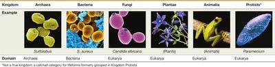
Types of Microbes
Microbe | Cell Type | Notes |
|---|---|---|
Bacteria | Prokaryotic | Unicellular; pathogenic and nonpathogenic |
Archaea | Prokaryotic | Unicellular; nonpathogenic; extremophiles |
Protists | Eukaryotic | Unicellular/multicellular; pathogenic and nonpathogenic |
Fungi | Eukaryotic | Unicellular/multicellular; pathogenic and nonpathogenic |
Helminths | Eukaryotic | Multicellular; parasitic worms |
Viruses | Nonliving | DNA or RNA genome; infects cells |
Prions | Nonliving | Infectious proteins; cause neurodegenerative diseases |
Historical Foundations of Microbiology
The Golden Age of Microbiology
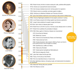
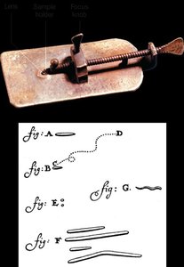
The period from 1850 to 1920 is known as the Golden Age of Microbiology, marked by major advances in microscopy, microbial isolation, and disease prevention. Key figures include Robert Hooke, Antonie van Leeuwenhoek, Louis Pasteur, and Robert Koch.
Spontaneous Generation vs. Biogenesis
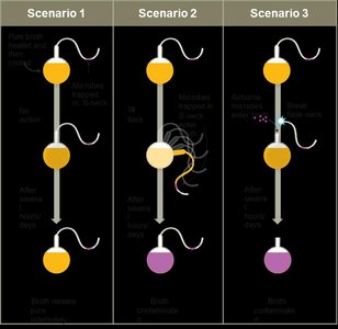
Early scientists debated whether life could arise from nonliving matter (spontaneous generation) or only from existing life (biogenesis). Experiments by Francesco Redi and Louis Pasteur provided evidence for biogenesis, disproving spontaneous generation.
Spontaneous generation: Life arises from nonliving matter.
Biogenesis: Life arises from pre-existing life.
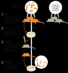
Germ Theory of Disease and Koch's Postulates
The germ theory of disease states that microbes are the cause of infectious diseases. Robert Koch developed a systematic method (Koch's postulates) to link specific microbes to specific diseases, revolutionizing medical microbiology.
Koch's Postulates: Criteria to establish a causative relationship between a microbe and a disease.
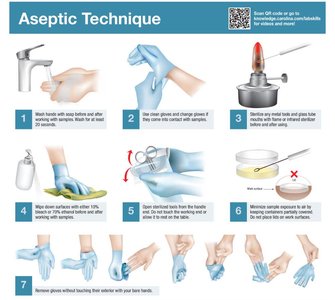
Aseptic Techniques and the Scientific Method
Aseptic Techniques
Aseptic techniques are procedures used to prevent contamination by unwanted microbes, crucial in healthcare and laboratory settings. Key contributors include Ignaz Semmelweis, Joseph Lister, and Florence Nightingale.
Hand washing
Wearing gloves
Sterilizing instruments
Decontaminating surfaces
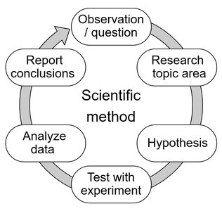
The Scientific Method
The scientific method is the foundation of scientific inquiry in microbiology. It involves making observations, forming hypotheses, conducting experiments, analyzing data, and drawing conclusions.
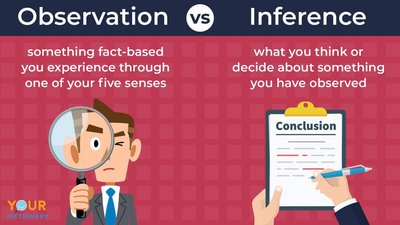
Observation: Gathering data using senses or instruments.
Hypothesis: A testable explanation for an observation.
Experiment: Testing the hypothesis under controlled conditions.
Conclusion: Interpretation of experimental results.
Taxonomy and Classification
Taxonomic Hierarchy
Taxonomy is the science of classifying organisms based on shared characteristics. The taxonomic hierarchy ranges from broad domains to specific species. The three domains are Bacteria, Archaea, and Eukarya.
Domain
Kingdom
Phylum
Class
Order
Family
Genus
Species
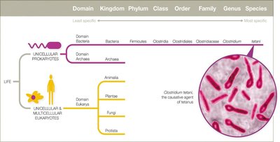
Scientific Nomenclature
Scientific names use binomial nomenclature: the genus (capitalized) and species (lowercase), both italicized (e.g., Escherichia coli). Strains are genetic variants within a species, often indicated by additional letters or numbers.
Host–Microbe Interactions and the Human Microbiome
Symbiosis and Pathogenicity
Microbes interact with hosts in various ways:
Parasitism: Microbe harms the host.
Mutualism: Both host and microbe benefit.
Commensalism: Microbe benefits; host is unaffected.
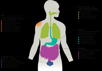
Normal microbiota (flora) are the collection of microbes that reside in and on the human body, often providing benefits such as immune training and vitamin production.
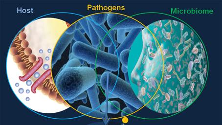
Microbiome and Human Health
The Human Microbiome Project aims to catalog all microbes associated with the human body. The microbiome influences digestion, immunity, and even mood. Disruptions (e.g., by antibiotics) can lead to infections by opportunistic pathogens.
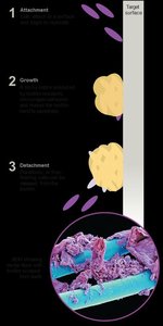
Biofilms
Biofilms are structured communities of microbes attached to surfaces and encased in a protective matrix. They are highly resistant to antibiotics and immune responses, and are implicated in many chronic infections.
Develop on teeth, catheters, water pipes, etc.
Account for 60–80% of human infections.
Microbial Cultivation and Laboratory Techniques
Growth Media and Culture Methods
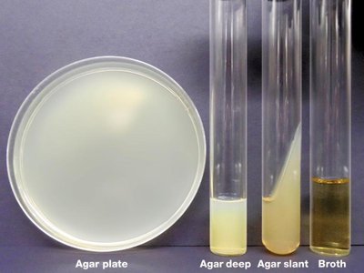
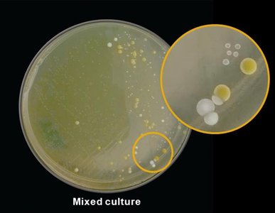
Microbes are grown in the laboratory using growth media, which may be liquid (broth) or solid (agar plates, slants, deeps). Julius Petri developed the petri dish for culturing microbes.
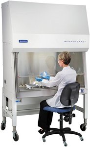
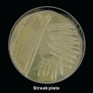
Aseptic Culture Techniques
Aseptic techniques are essential for isolating pure cultures and preventing contamination. Biological safety cabinets and streak plate methods are commonly used.
Microscopy and Staining Techniques
Staining Methods
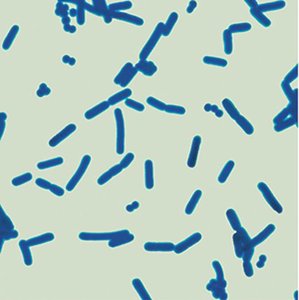
Stains increase contrast in microscopic samples. Basic dyes (e.g., methylene blue, crystal violet) stain cells, while acidic dyes (e.g., nigrosin) stain the background. Mordants (e.g., iodine) help fix dyes to cells.
Simple stains: Use one dye to reveal cell shape and arrangement.
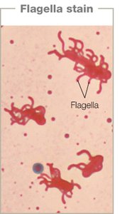
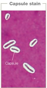
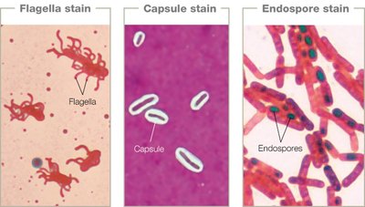
Structural stains: Highlight specific structures (flagella, capsules, endospores).
Differential stains: Distinguish between cell types (Gram stain, acid-fast stain).

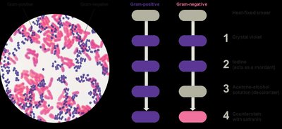
Gram Stain
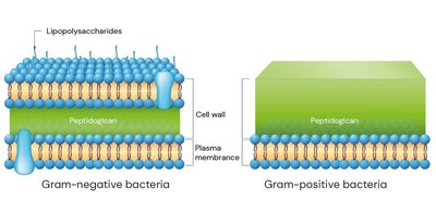
The Gram stain differentiates bacteria based on cell wall structure:
Gram-positive: Thick peptidoglycan layer, stains purple.
Gram-negative: Thin peptidoglycan layer, outer membrane, stains pink.
Steps: Crystal violet (primary stain) → Iodine (mordant) → Alcohol (decolorizer) → Safranin (counterstain).
Acid-Fast Stain
Acid-fast staining distinguishes bacteria with waxy cell walls (rich in mycolic acid), such as Mycobacterium species. Acid-fast cells retain the red dye after acid wash; non–acid-fast cells do not.
Microscopy Techniques
Light Microscopy
Light microscopes use visible light and lenses to magnify specimens. The compound light microscope is the most common type, with magnification up to 1,500x and resolution of about 200 nm.
Bright field: Standard illumination; stained or naturally colored samples.
Dark field: Illuminates sample with hollow cone of light; unstained specimens appear bright on dark background.
Phase contrast: Enhances contrast in unstained specimens using phase shifts in light.
Differential interference contrast: Uses polarized light for 3D-like images.
Electron Microscopy
Electron microscopes use electron beams for much higher resolution (up to 0.2 nm). Two main types:
Transmission Electron Microscopy (TEM): Electrons pass through thin specimens; reveals internal structures (2D images).
Scanning Electron Microscopy (SEM): Electrons scan the surface; reveals surface details (3D images).
Fluorescence and Probe Microscopy
Fluorescence microscopy uses dyes that emit light under UV illumination, allowing detection of specific molecules or structures. Probe microscopy (e.g., atomic force microscopy) can visualize surfaces at the atomic level.
Clinical Applications and Case Studies
Cholera Case Study
Cholera, caused by Vibrio cholerae, is a waterborne disease. Historical and modern studies illustrate the importance of proper sanitation, microbial identification, and understanding host-microbe interactions in disease prevention and management.
Summary Table: Key Differences in Microbial Classification
Feature | Prokaryotes | Eukaryotes | Viruses/Prions |
|---|---|---|---|
Cell Type | Prokaryotic | Eukaryotic | Non-cellular |
Examples | Bacteria, Archaea | Fungi, Protists, Helminths | Viruses, Prions |
Genetic Material | DNA (circular) | DNA (linear) | DNA or RNA (viruses); protein (prions) |
Reproduction | Binary fission | Mitosis/meiosis | Requires host cell (viruses); misfolding (prions) |
