 Back
BackIntroduction to Microbiology: Core Concepts and Methods
Study Guide - Smart Notes
 Tailored notes based on your materials, expanded with key definitions, examples, and context.
Introduction to Microbiology
Definition and Scope
Microbiology is the scientific study of microorganisms, or microbes, which are typically too small to be seen with the naked eye. The field encompasses both cellular, living microorganisms (such as bacteria, archaea, fungi, protists, and helminths) and nonliving entities (such as viruses and prions). Microbes inhabit nearly every environment on Earth, from deep-sea trenches to glaciers, and constitute at least half of Earth's biomass.
Microbe: A microscopic organism, which may be unicellular or multicellular.
Pathogen: A microbe that causes disease; less than 1% of microbes are pathogenic.
Opportunistic pathogen: Causes disease only in weakened hosts.
Applications: Microbiology is central to healthcare, agriculture, industry, and environmental sciences. Humans rely on microbes for food production, medication synthesis, and environmental remediation.
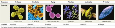
Types of Microbes
Microbe | Cell Type | Notes |
|---|---|---|
Bacteria | Prokaryotic | Unicellular; pathogenic and nonpathogenic |
Archaea | Prokaryotic | Unicellular; nonpathogenic; extremophiles |
Protists | Eukaryotic | Unicellular/multicellular; pathogenic and nonpathogenic |
Fungi | Eukaryotic | Unicellular/multicellular; pathogenic and nonpathogenic |
Helminths | Eukaryotic | Multicellular; parasitic worms |
Viruses | Nonliving | DNA or RNA genome; infects cells |
Prions | Nonliving | Infectious proteins; cause neurodegenerative diseases |
Historical Foundations of Microbiology
The Golden Age of Microbiology
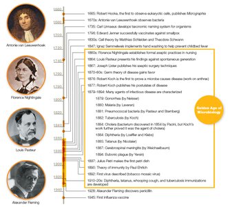
The period from 1850 to 1920 is known as the Golden Age of Microbiology, marked by major advances in microscopy, microbial isolation, and disease prevention. Key figures include Robert Hooke, Antonie van Leeuwenhoek, Louis Pasteur, and Robert Koch.
Spontaneous Generation vs. Biogenesis
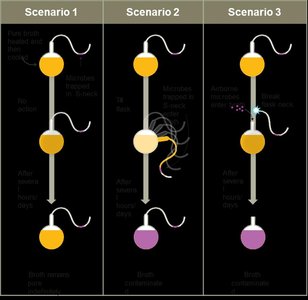
Early scientists debated whether life arose spontaneously from nonliving matter (spontaneous generation) or from existing life (biogenesis). Experiments by Francesco Redi and Louis Pasteur provided evidence for biogenesis, disproving spontaneous generation.
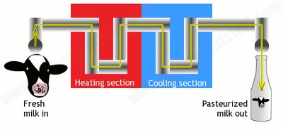
Pasteurization: A process developed by Pasteur to kill microbes in food and beverages by heating to 50–60°C.
Germ Theory of Disease and Koch's Postulates
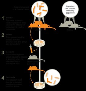
The germ theory of disease posits that microbes cause infectious diseases. Robert Koch developed a systematic method (Koch's postulates) to link specific microbes to specific diseases, using isolation, cultivation, and infection experiments.
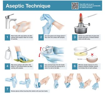
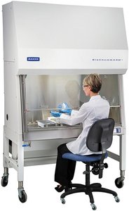
Aseptic Techniques and Infection Control
Development of Aseptic Techniques
Medical professionals such as Ignaz Semmelweis, Joseph Lister, and Florence Nightingale pioneered aseptic techniques to prevent healthcare-associated infections (HAIs). These include hand washing, sterilizing instruments, and decontaminating surfaces.
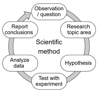
The Scientific Method in Microbiology
Principles and Application
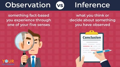
The scientific method is the foundation of microbiological research. It involves formulating a question, proposing a hypothesis, collecting and analyzing data, and drawing conclusions. Distinguishing between observations (data) and conclusions (interpretations) is critical for accurate scientific inquiry.
Scientific Laws vs. Theories
Law: A precise statement or mathematical formula predicting a specific occurrence.
Theory: A hypothesis supported by extensive evidence, explaining how and why phenomena occur.
Both laws and theories are subject to revision as new evidence emerges.
Taxonomy and Classification of Microbes
Taxonomic Hierarchy
Taxonomy is the science of classifying organisms based on shared characteristics. The hierarchy includes Domain, Kingdom, Phylum, Class, Order, Family, Genus, and Species. Carl Linnaeus established the binomial nomenclature system for naming organisms.
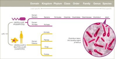
Species and Strains
Eukaryotic species: Organisms that can sexually reproduce together.
Prokaryotic species: Cells with at least 70% DNA similarity and 97% 16S rRNA sequence similarity.
Strain: Genetic variants within a species, often denoted by numbers or letters (e.g., E. coli K-12).
Host–Microbe Interactions
Symbiosis and Pathogenicity
Microbes and hosts can have various symbiotic relationships:
Parasitism: Microbe harms the host (e.g., pathogens).
Mutualism: Both benefit.
Commensalism: One benefits, the other is unaffected.
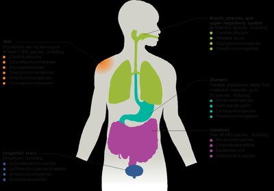
Normal microbiota (flora) are the collection of microbes residing in and on the human body, often providing mutualistic benefits such as immune training and vitamin production.
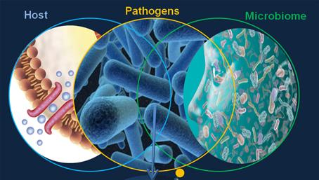
Microbiome and Human Health
The Human Microbiome Project aims to characterize all microbes associated with humans. The microbiome influences immunity, digestion, and even mood. Disruptions (e.g., by antibiotics) can lead to infections by opportunistic pathogens.
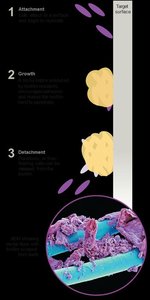
Biofilms
Formation and Significance
Biofilms are structured communities of microbes attached to surfaces and encased in a protective matrix. They are highly resistant to antibiotics and immune responses, and are implicated in 60–80% of human infections.
Microbial Culture and Laboratory Techniques
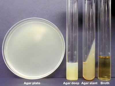
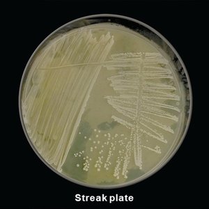
Growth Media and Culture Methods
Growth media provide nutrients for microbial growth in the lab. Media types include broths, plates, slants, and deeps. Agar is a common solidifying agent. Aseptic techniques are essential to prevent contamination.
Microscopy and Staining Techniques
Staining Methods
Stains increase contrast for microscopic observation. Basic dyes (e.g., methylene blue, crystal violet) stain cells, while acidic dyes stain backgrounds. Mordants (e.g., iodine) fix dyes to specimens. Staining techniques include simple, structural, and differential stains.
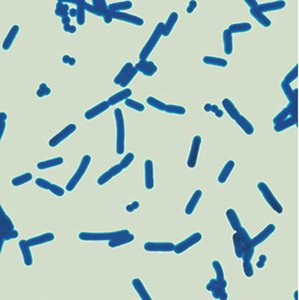
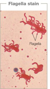
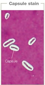
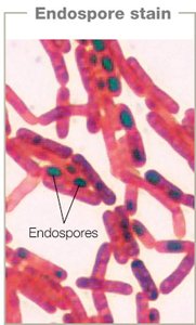
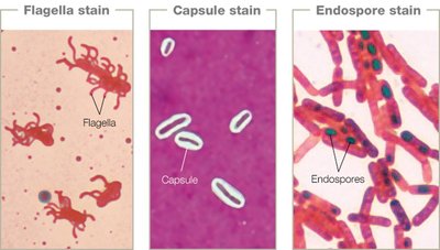
Structural Stains
Flagella stain: Visualizes bacterial flagella.
Capsule stain: Highlights bacterial capsules as clear halos.
Endospore stain: Differentiates endospores from vegetative cells.
Differential Stains: Gram and Acid-Fast
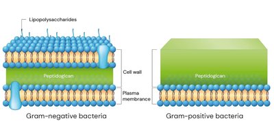
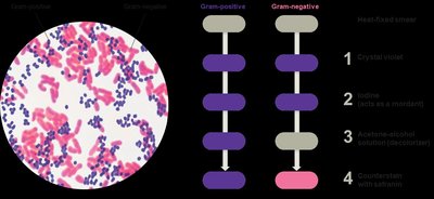
Gram staining differentiates bacteria based on cell wall structure:
Gram-positive: Thick peptidoglycan, stains purple.
Gram-negative: Thin peptidoglycan, outer membrane, stains pink.
Acid-fast staining identifies bacteria with waxy cell walls (e.g., Mycobacterium species). Acid-fast cells retain red dye after acid wash; non–acid-fast cells do not.
Microscopy Techniques
Light Microscopy
Light microscopes use visible light and lenses to magnify specimens. Types include bright field, dark field, phase contrast, and differential interference contrast. The compound light microscope is most common, with magnifications up to 1,500x and resolution of about 200 nm.
Electron Microscopy
Electron microscopes use electron beams for much higher resolution (up to 0.2 nm). Types include transmission electron microscopy (TEM) for internal structures and scanning electron microscopy (SEM) for surface details.
Fluorescence and Probe Microscopy
Fluorescence microscopy uses dyes that emit light under UV illumination, allowing sensitive detection of specific molecules or structures. Probe microscopy (e.g., atomic force microscopy) can visualize surfaces at the atomic level.
Clinical Application: Cholera Case Study
Summary and Key Concepts
Cholera is caused by Vibrio cholerae, a waterborne bacterium.
Proper sewage management reduces cholera by preventing water contamination.
Symbiotic relationships can be mutualistic (e.g., V. cholerae and copepods) or pathogenic (in humans).
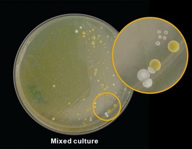
Microbial strains can differ in survival and pathogenicity based on colony morphology (e.g., rugose vs. smooth).
